
Cardiovascular Nursing & Acute Coronary Syndrome
In this chapterفي هذا الفصل
Key terms (12)المصطلحات الأساسية
Chapter outline (40 sections)مخطط الفصل
- Learning Objectivesأهداف التعلّم
- 1.1 · Focused Cardiac Assessment١٫١ · التقييم القلبي المركَّز
- 1.2 · Conduction and Coronary Anatomy١٫٢ · التوصيل وتشريح الشرايين التاجية
- 1.3 · Heart Sounds١٫٣ · أصوات القلب
- 1.4 · The ECG and the Arrest Rhythms١٫٤ · تخطيط القلب ونظم التوقّف
- 1.5 · Cardiac Biomarkers١٫٥ · واسمات القلب
- 1.6 · Non-Invasive Cardiac Investigations١٫٦ · فحوصات القلب غير الباضعة
- 1.7 · ECG Leads and the Dangerous Rhythms١٫٧ · اشتقاقات التخطيط والنظم الخطرة
- 1.8 · Cardiac Catheterisation Care١٫٨ · رعاية قسطرة القلب
- 1.9 · Cardiovascular Risk and Prevention١٫٩ · الخطر القلبي الوعائي والوقاية
- 1.10 · Angina١٫١٠ · الذبحة الصدرية
- 1.11 · Acute Coronary Syndrome: Presentation١٫١١ · المتلازمة التاجية الحادّة: التظاهر
- 1.12 · Acute Coronary Syndrome: Immediate Management١٫١٢ · المتلازمة التاجية الحادّة: التدبير الفوري
- 1.13 · After the Infarct: Complications١٫١٣ · بعد الاحتشاء: المضاعفات
- 1.14 · Reperfusion, Thrombolysis and Bleeding Precautions١٫١٤ · إعادة التروية وحلّ الخثرة واحتياطات النزف
- 1.15 · Heart Failure: Left and Right١٫١٥ · قصور القلب: الأيسر والأيمن
- 1.16 · Managing Heart Failure١٫١٦ · تدبير قصور القلب
- 1.17 · Acute Pulmonary Oedema١٫١٧ · الوذمة الرئوية الحادّة
- 1.18 · Atrial Fibrillation١٫١٨ · الرجفان الأذيني
- 1.19 · Grading Heart Failure and Oedema١٫١٩ · تدريج قصور القلب والوذمة
- 1.20 · Bradycardia and Heart Block١٫٢٠ · بطء النبض وحصار القلب
- 1.21 · Tachyarrhythmias and Cardioversion١٫٢١ · التسرّعات القلبية وتقويم النظم
- 1.22 · Cardiac Arrest and After١٫٢٢ · توقّف القلب وما بعده
- 1.23 · Pacemakers and Implantable Defibrillators١٫٢٣ · نواظم الخطى ومزيلات الرجفان المزروعة
- 1.24 · Pericarditis and Cardiac Tamponade١٫٢٤ · التهاب التامور واندحاس القلب
- 1.25 · Valvular Disease١٫٢٥ · أمراض الصمّامات
- 1.26 · Hypertension and Hypertensive Emergency١٫٢٦ · ارتفاع ضغط الدم وطارئ ارتفاع ضغط الدم
- 1.27 · Arterial and Venous Limb Disease١٫٢٧ · مرض الطرف الشرياني والوريدي
- 1.28 · Venous Thromboembolism and Aortic Emergencies١٫٢٨ · الانصمام الخثاري الوريدي وطوارئ الأبهر
- 1.29 · Cardiac Surgery and Haemodynamic Monitoring١٫٢٩ · جراحة القلب ومراقبة الديناميكا الدموية
- 1.30 · Other Vascular Conditions١٫٣٠ · حالات وعائية أخرى
- 1.31 · Shock١٫٣١ · الصدمة
- 1.32 · Cardiomyopathy, Rheumatic Disease and Prophylaxis١٫٣٢ · أمراض عضلة القلب، والمرض الروماتيزمي، والوقاية
- 1.33 · Antiplatelets and Anticoagulants١٫٣٣ · مضادّات الصفيحات ومضادّات التخثّر
- 1.34 · Direct Oral Anticoagulants and Supportive Drugs١٫٣٤ · مضادّات التخثّر الفموية المباشرة والأدوية الداعمة
- 1.35 · Preventing Venous Thromboembolism١٫٣٥ · الوقاية من الانصمام الخثاري الوريدي
- 1.36 · Beta-Blockers, ACE Inhibitors and Calcium Channel Blockers١٫٣٦ · حاصرات بيتا ومثبّطات الإنزيم المحوِّل للأنجيوتنسين وحاصرات الكالسيوم
- 1.37 · Nitrates١٫٣٧ · النترات
- 1.38 · Digoxin, Diuretics and Statins١٫٣٨ · الديجوكسين والمدرّات والستاتينات
- 1.39 · Cardiovascular Care in the Saudi Setting١٫٣٩ · الرعاية القلبية الوعائية في السياق السعودي
Start learningبدء التعلّم Learning objectivesأهداف التعلّم Test yourselfالاختبار الذاتي
Learning Objectivesأهداف التعلّم
1.1 · Focused Cardiac Assessment١٫١ · التقييم القلبي المركَّز
Start with the history: the character, site, radiation, timing and triggers of any chest pain, breathlessness and what brings it on, orthopnoea and how many pillows he sleeps on, paroxysmal nocturnal dyspnoea, palpitations, syncope, oedema, and exercise tolerance in functional terms — how far he can walk, how many stairs.
يُبدأ بالقصّة: طبيعة ألم الصدر وموقعه وانتشاره وتوقيته ومحفّزاته، وضيق النفس وما يُثيره، وضيق التنفّس الاضطجاعي وعدد الوسائد التي ينام عليها، وضيق النفس الليلي الانتيابي، والخفقان، والإغماء، والوذمة، وتحمّل الجهد بمصطلحات وظيفية — كم يمشي وكم درجة يصعد.
Inspect and palpate: colour and perfusion, capillary refill, peripheral and central cyanosis, jugular venous pressure, peripheral oedema and where it collects, the apex beat, and all peripheral pulses bilaterally and compared. A difference between the two arms is itself a finding.
يُفحَص بالنظر والجسّ: اللون والتروية، وعود الشعيرات، والزرقة الطرفية والمركزية، والضغط الوريدي الوداجي، والوذمة الطرفية وأين تتجمّع، وضربة القمّة، وكلّ النبضات الطرفية في الجانبين مع المقارنة. والفرق بين الذراعين بحدّ ذاته علامة سريرية.
The five auscultation areas: aortic at the second right intercostal space, pulmonic at the second left, Erb's point at the third left, tricuspid at the fourth left, and mitral or apex at the fifth left in the midclavicular line — which is where the apical pulse is counted, as Chapter 04 of Part 01 sets out.
مواضع الإصغاء الخمسة: الأبهري عند المسافة الوربية الثانية اليمنى، والرئوي عند الثانية اليسرى، ونقطة إرب عند الثالثة اليسرى، وثلاثي الشرف عند الرابعة اليسرى، والتاجي أو القمّة عند الخامسة اليسرى على خطّ منتصف الترقوة — وهو الموضع الذي يُعدّ فيه النبض القمّي كما يبيّن الفصل الرابع من الجزء الأول.
1.2 · Conduction and Coronary Anatomy١٫٢ · التوصيل وتشريح الشرايين التاجية
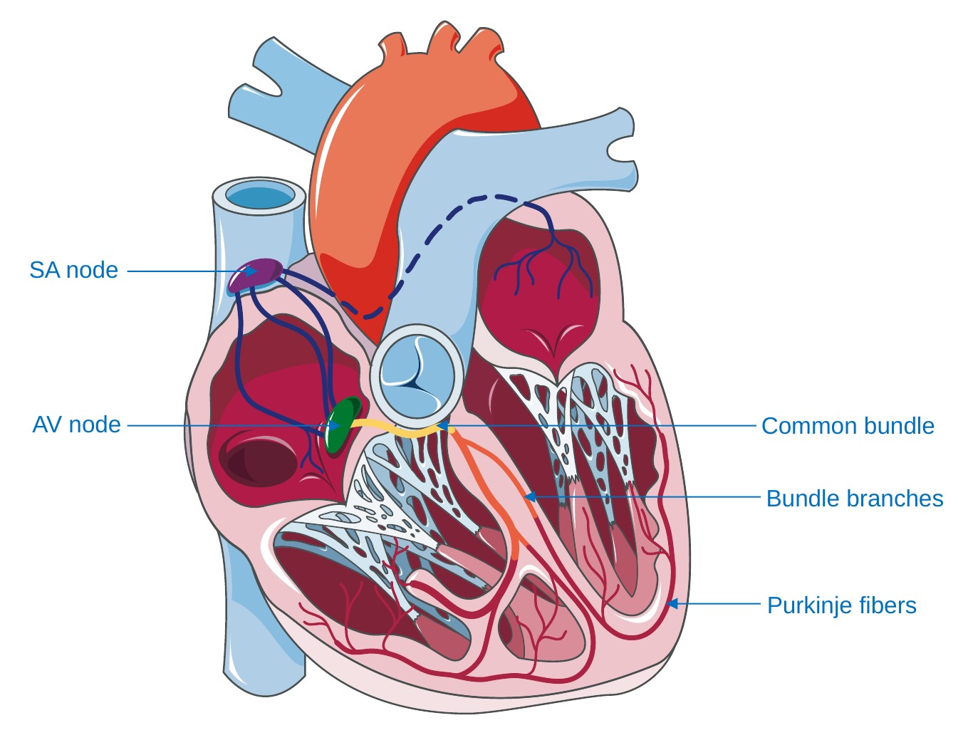
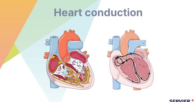
The impulse begins at the sinoatrial node, the natural pacemaker firing at about 60 to 100 a minute, travels through the atria to the atrioventricular node, which delays it so the atria can empty, then down the bundle of His, the bundle branches and the Purkinje fibres into the ventricles. If the sinoatrial node fails, the atrioventricular node takes over at about 40 to 60, and the ventricles at about 20 to 40 — which is why an escape rhythm is slow.
تبدأ السيالة عند العقدة الجيبية الأذينية، الناظم الطبيعي الذي يُطلق نحو ٦٠ إلى ١٠٠ في الدقيقة، وتنتقل عبر الأذينين إلى العقدة الأذينية البطينية التي تؤخّرها ليفرغ الأذينان، ثم تنزل عبر حزمة هيس وغصنيها وألياف بركنجي إلى البطينين. وإن فشلت العقدة الجيبية تولّت الأذينية البطينية بنحو ٤٠ إلى ٦٠، والبطينان بنحو ٢٠ إلى ٤٠ — ولهذا يكون نظم الهروب بطيئاً.
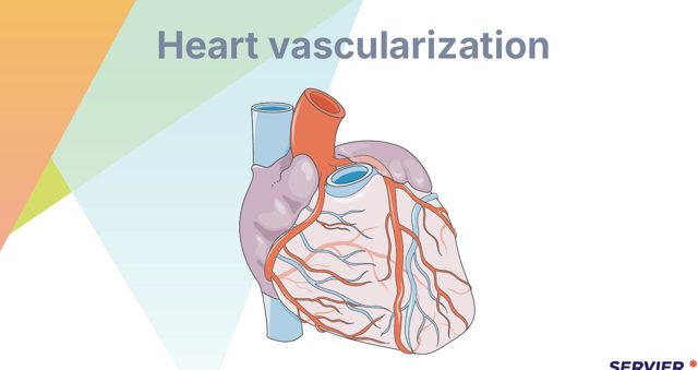
Three coronary arteries supply the muscle and each maps to a wall and to a set of leads. The left anterior descending supplies the anterior wall and septum — leads V1 to V4. The circumflex supplies the lateral wall — leads I, aVL, V5 and V6. The right coronary artery supplies the inferior wall — leads II, III and aVF — and in most people it also supplies the sinoatrial and atrioventricular nodes.
ثلاثة شرايين تاجية تغذّي العضلة ويرتبط كلّ منها بجدار ومجموعة اشتقاقات. فالأمامي النازل الأيسر يغذّي الجدار الأمامي والحاجز — الاشتقاقات V1 إلى V4. والمنعطف يغذّي الجدار الوحشي — الاشتقاقات I وaVL وV5 وV6. والشريان التاجي الأيمن يغذّي الجدار السفلي — الاشتقاقات II وIII وaVF — وهو لدى معظم الناس يغذّي أيضاً العقدتين الجيبية والأذينية البطينية.
That last point explains something the exam relies on: an inferior infarct commonly causes bradycardia and heart block, because the artery that has occluded is also the one supplying the conduction nodes. A patient with ST elevation in II, III and aVF who becomes bradycardic is behaving exactly as the anatomy predicts.
تلك النقطة الأخيرة تفسّر ما يعتمد عليه الامتحان: فالاحتشاء السفلي يسبّب عادةً بطء نبض وحصار قلب، لأن الشريان المنسدّ هو نفسه الذي يغذّي عقد التوصيل. والمريض بارتفاع ST في II وIII وaVF الذي يصير بطيء النبض يتصرّف تماماً كما يتنبّأ التشريح.
1.3 · Heart Sounds١٫٣ · أصوات القلب
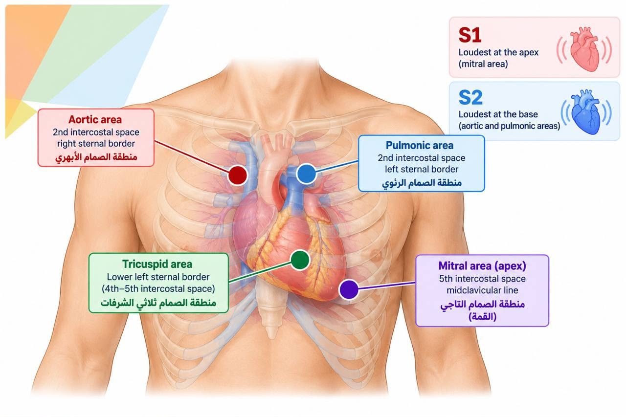
S1 is the closure of the mitral and tricuspid valves at the start of systole, loudest at the apex. S2 is the closure of the aortic and pulmonic valves at the start of diastole, loudest at the base.
الصوت الأول هو انغلاق الصمّامين التاجي وثلاثي الشرف في بداية الانقباض، وأعلى ما يكون عند القمّة. والصوت الثاني انغلاق الصمّامين الأبهري والرئوي في بداية الانبساط، وأعلى ما يكون عند القاعدة.
S3 — the ventricular gallop — occurs in early diastole as blood rushes into a ventricle that is already full. It can be normal in children, young adults and pregnancy, but in an older adult it indicates volume overload and is a hallmark of heart failure.
الصوت الثالث — الخبب البطيني — يحدث في بداية الانبساط مع اندفاع الدم إلى بطين ممتلئ أصلاً. وقد يكون طبيعياً لدى الأطفال والشباب والحوامل، لكنه لدى كبير السنّ يدلّ على فرط الحمل الحجمي وهو علامة مميّزة لقصور القلب.
S4 — the atrial gallop — occurs in late diastole as the atrium contracts against a stiff, non-compliant ventricle. It is heard in hypertension, aortic stenosis, ischaemia and ventricular hypertrophy.
الصوت الرابع — الخبب الأذيني — يحدث في نهاية الانبساط مع انقباض الأذين على بطين متيبّس قليل المطاوعة. ويُسمع في ارتفاع ضغط الدم وتضيّق الأبهر ونقص التروية وضخامة البطين.
A pericardial friction rub is a scratchy, grating sound heard best with the diaphragm at the left sternal border with the patient sitting forward and holding his breath out. It is the sound of inflamed pericardial surfaces rubbing together.
احتكاك التامور صوت خشن كالحكّ يُسمع على أفضل وجه بغشاء السمّاعة عند حافّة القصّ اليسرى والمريض مائل للأمام وحابس زفيره. وهو صوت احتكاك سطحي التامور الملتهبين.
1.4 · The ECG and the Arrest Rhythms١٫٤ · تخطيط القلب ونظم التوقّف
In suspected acute coronary syndrome a 12-lead ECG is obtained within 10 minutes of arrival, and that is a nursing responsibility rather than something to wait for. ST elevation in contiguous leads (or an equivalent pattern, such as a left bundle branch block that meets the Sgarbossa criteria) indicates a STEMI and triggers the reperfusion pathway.
في المتلازمة التاجية الحادّة المشتبهة يُؤخذ تخطيط قلب بـ١٢ اشتقاقاً خلال عشر دقائق من الوصول، وهي مسؤولية تمريضية وليست أمراً يُنتظر. وارتفاع ST في اشتقاقات متجاورة (أو نمط معادل له، كحصار غصن أيسر يستوفي معايير Sgarbossa) يدلّ على احتشاء بارتفاع ST ويُفعّل مسار إعادة التروية.
The four arrest rhythms divide by whether a shock will help. Shockable: ventricular fibrillation and pulseless ventricular tachycardia — defibrillate. Non-shockable: asystole and pulseless electrical activity — high-quality compressions and adrenaline, and look for the reversible causes.
نظم التوقّف الأربعة تنقسم بحسب ما إذا كانت الصدمة ستنفع. القابلة للصدمة: الرجفان البطيني والتسرّع البطيني بلا نبض — تُجرى إزالة الرجفان. وغير القابلة: انعدام الانقباض والنشاط الكهربائي بلا نبض — ضغطات عالية الجودة وأدرينالين، والبحث عن الأسباب القابلة للعكس.
Electrolytes change the trace and the exam asks about them. Hyperkalaemia: tall peaked T waves, then a widening QRS, then a sine wave and arrest. Hypokalaemia: flattened T waves, ST depression and a U wave. Hypokalaemia also potentiates digoxin toxicity, which is covered later in this chapter.
الشوارد تغيّر التخطيط والاختبار يسأل عنها. فرط البوتاسيوم: موجات T طويلة مدبّبة، ثم اتّساع مركّب QRS، ثم موجة جيبية وتوقّف. ونقص البوتاسيوم: تسطّح موجات T وانخفاض ST وموجة U. ونقص البوتاسيوم يزيد أيضاً سمّية الديجوكسين المشروحة لاحقاً في هذا الفصل.
1.5 · Cardiac Biomarkers١٫٥ · واسمات القلب
CK-MB rises at about 4 to 6 hours, peaks around 18 to 24 hours and returns to normal within 48 to 72 hours, which makes it the best alternative for detecting re-infarction when a troponin assay is not available. Myoglobin rises earliest but is not specific to cardiac muscle.
الكرياتين كيناز MB يرتفع عند نحو أربع إلى ستّ ساعات، ويبلغ الذروة نحو ١٨ إلى ٢٤ ساعة، ويعود طبيعياً خلال ٤٨ إلى ٧٢ ساعة، وهو ما يجعله أفضل بديل لكشف الاحتشاء المتكرّر حين يتعذّر توافر فحص التروبونين. والميوغلوبين يرتفع أبكر لكنه غير نوعي للعضلة القلبية.
BNP and NT-proBNP are released when the ventricle is stretched, so they are markers of heart failure rather than of infarction, and they help distinguish a cardiac cause of breathlessness from a respiratory one.
الببتيد الدماغي المدرّ للصوديوم (BNP) وNT-proBNP يُطلَقان عند تمدّد البطين، فهما واسمان لقصور القلب لا للاحتشاء، ويساعدان على تمييز السبب القلبي لضيق النفس من السبب التنفّسي.
1.6 · Non-Invasive Cardiac Investigations١٫٦ · فحوصات القلب غير الباضعة
A single early troponin does not exclude infarction, because the level takes hours to rise. Blood is taken on arrival and repeated at the interval the local protocol specifies, and it is the change between the two samples as much as the absolute value that makes the diagnosis. A patient with a normal first troponin is not cleared until the second is back.
التروبونين المبكّر الواحد لا ينفي الاحتشاء، فالمستوى يحتاج ساعات ليرتفع. فيُسحب الدم عند الوصول ويُعاد بالفاصل الذي يحدّده البروتوكول المحلّي، والتغيّر بين العيّنتين يصنع التشخيص بقدر القيمة المطلقة. والمريض بتروبونين أول طبيعي لا يُخلى سبيله حتى تعود العيّنة الثانية.
Exercise stress testing looks for ischaemia that appears only under load. Preparation: nothing by mouth for a few hours, no caffeine, comfortable shoes and loose clothing, and clarify with the prescriber which cardiac drugs — beta-blockers in particular — are to be held, since they blunt the heart rate response. The test is stopped for chest pain, significant ST change, arrhythmia, a falling blood pressure or exhaustion.
اختبار الجهد يبحث عن نقص تروية لا يظهر إلا تحت الحمل. والتحضير: صيام بضع ساعات، ولا كافيين، وحذاء مريح وملابس فضفاضة، وتوضيح مع الطبيب أيّ أدوية قلبية — وحاصرات بيتا خاصّةً — تُحجَب، فهي تخمد استجابة النبض. ويُوقَف الاختبار لألم صدر أو تغيّر ST ذي دلالة أو اضطراب نظم أو هبوط ضغط أو إنهاك.
Echocardiography gives the ejection fraction — the proportion of blood the left ventricle expels with each beat. A normal fraction is roughly 55 to 70%; a value of 40% or below defines heart failure with reduced ejection fraction. A Holter monitor records the rhythm over 24 hours or longer, and the patient keeps a symptom diary so any palpitations can be matched to the trace.
تخطيط صدى القلب يعطي الكسر القذفي — نسبة الدم التي يقذفها البطين الأيسر مع كلّ نبضة. والكسر الطبيعي نحو ٥٥ إلى ٧٠٪؛ والقيمة ٤٠٪ أو أقلّ تُعرّف قصور القلب منخفض الكسر القذفي. وجهاز هولتر يسجّل النظم ٢٤ ساعة أو أكثر، ويحتفظ المريض بمذكّرة أعراض لمطابقة أيّ خفقان بالتخطيط.
1.7 · ECG Leads and the Dangerous Rhythms١٫٧ · اشتقاقات التخطيط والنظم الخطرة
Chest lead placement must be ACCURATE, because a DISPLACED LEAD CHANGES THE TRACE and can CREATE or HIDE an infarct.
يجب أن يكون وضع اشتقاقات الصدر دقيقاً، لأن القطب المزاح يغيّر التخطيط وقد يخلق احتشاءً أو يُخفيه.
- WIDE, BIZARRE complexes with NO preceding P wave
- Occasional ones are COMMON and often BENIGN
- Concerning when FREQUENT, MULTIFOCAL, in RUNS
- or falling on the T wave (R-on-T)
- Check POTASSIUM and MAGNESIUM
- POLYMORPHIC VT — complexes TWIST around the baseline
- Arises from a PROLONGED QT INTERVAL
- Congenital, from ELECTROLYTES, or from DRUGS
- Treated with MAGNESIUM
- And the drug list is reviewed — several antiemetics, antipsychotics and antibiotics prolong the QT
Right ventricular infarction accompanies about 40% of inferior infarcts (up to half) and is confirmed with right-sided chest leads. It matters enormously for nursing because the right ventricle becomes preload dependent: nitrates, diuretics and morphine can cause profound hypotension in these patients, and the treatment is fluid rather than the vasodilator that would be right in an ordinary infarct. A patient who becomes severely hypotensive after a nitrate for an inferior infarct has probably had a right ventricular infarct.
احتشاء البطين الأيمن يرافق نحو ٤٠٪ من الاحتشاءات السفلية (وحتى النصف) ويُؤكَّد باشتقاقات صدرية يمنى. وهو يهمّ التمريض كثيراً لأن البطين الأيمن يصير معتمداً على الحمل القبلي: فالنترات والمدرّات والمورفين قد تسبّب هبوط ضغط عميقاً لدى هؤلاء المرضى، والعلاج سوائل لا الموسّع الوعائي الذي يصحّ في احتشاء عادي. والمريض الذي يهبط ضغطه بشدّة بعد نترات لاحتشاء سفلي أُصيب على الأرجح باحتشاء بطين أيمن.
Premature ventricular contractions are wide, bizarre complexes with no preceding P wave. Occasional ones are common and often benign; they become concerning when they are frequent, multifocal, occur in runs, or fall on the T wave of the preceding beat, since that can trigger ventricular tachycardia. Look for the treatable causes — hypokalaemia, hypomagnesaemia, hypoxia, ischaemia, caffeine and drug toxicity.
الانقباضات البطينية المبكّرة مركّبات عريضة شاذّة بلا موجة P سابقة. والعارضة منها شائعة وحميدة غالباً؛ وتصير مقلقة حين تكون متكرّرة أو متعدّدة البؤر أو في سلاسل أو تقع على موجة T للنبضة السابقة، فذلك قد يُحدث تسرّعاً بطينياً. ويُبحث عن الأسباب القابلة للعلاج — نقص البوتاسيوم والمغنيسيوم ونقص الأكسجة ونقص التروية والكافيين والسمّية الدوائية.
Torsades de pointes is a polymorphic ventricular tachycardia in which the complexes appear to twist around the baseline. It arises from a prolonged QT interval, whether congenital, from electrolyte disturbance or from QT-prolonging drugs. The treatment that distinguishes it is intravenous magnesium sulfate, alongside correcting the electrolytes and stopping the offending drug; if the patient is pulseless it is defibrillated as for any pulseless ventricular rhythm.
تورساد دو بوانت تسرّع بطيني متعدّد الأشكال تبدو مركّباته ملتفّة حول خطّ الأساس. وينشأ من إطالة فترة QT، خلقية كانت أو من اضطراب شاردي أو من أدوية مطيلة لـQT. والعلاج الذي يميّزه كبريتات المغنيسيوم وريدياً، إلى جانب تصحيح الشوارد وإيقاف الدواء المسبّب؛ وإن كان المريض بلا نبض فتُجرى إزالة الرجفان كما في أيّ نظم بطيني بلا نبض.
1.8 · Cardiac Catheterisation Care١٫٨ · رعاية قسطرة القلب
Before: confirm consent, check for any previous reaction to contrast media (a shellfish or ‘iodine’ allergy does not predict one), check renal function since the contrast is nephrotoxic, confirm fasting status, record baseline observations and mark the distal pulses so they can be found again quickly afterwards. Ask about metformin, which is often withheld around contrast.
قبلها: يُتأكَّد من الحصول على الموافقة، ويُتحقَّق من أيّ تفاعل سابق مع الصبغة (فحساسية المحار أو «اليود» لا تتنبّأ بتفاعل معها)، وتُفحَص وظيفة الكلى فالصبغة سامّة كلوياً، وتُؤكَّد حالة الصيام، وتُسجَّل القياسات الأساسية، وتُعلَّم النبضات البعيدة ليسهل إيجادها بعدها. ويُسأل عن الميتفورمين الذي يُحجَب غالباً حول الصبغة.
Afterwards, the first priority is the limb distal to the puncture site and the site itself: check the pulse, colour, temperature, sensation and movement, and inspect for bleeding or an expanding haematoma. Keep the limb straight and the patient on bed rest for the period policy specifies, and encourage fluids to clear the contrast.
بعدها، الأولوية الأولى هي الطرف البعيد عن موقع الوخز والموقع نفسه: يُفحص النبض واللون والحرارة والإحساس والحركة، ويُفحص بحثاً عن نزف أو ورم دموي متمدّد. ويُبقى الطرف مستقيماً والمريض على راحة فراش للمدّة التي تحدّدها السياسة، ويُشجَّع على تناول السوائل لطرح الصبغة.
Watch for the complication that is easily missed: retroperitoneal bleeding after a femoral approach presents with back or flank pain, hypotension and a falling haemoglobin with no visible bleeding at the site. A patient who becomes hypotensive after a femoral cath and has back pain is bleeding until proved otherwise.
يُراقَب ظهور المضاعفة سهلة الإغفال: فالنزف خلف الصفاق بعد المدخل الفخذي يتظاهر بألم ظهر أو خاصرة وهبوط ضغط وانخفاض الخضاب دون نزف ظاهر في الموقع. والمريض الذي يهبط ضغطه بعد قسطرة فخذية ولديه ألم ظهر ينزف حتى يثبت العكس.
1.9 · Cardiovascular Risk and Prevention١٫٩ · الخطر القلبي الوعائي والوقاية
Non-modifiable risk factors are age, male sex — with women's risk rising after the menopause — a family history of premature coronary disease, and ethnicity. They cannot be changed, but they change how aggressively the modifiable factors are treated.
عوامل الخطر غير القابلة للتعديل هي العمر والجنس الذكري — مع ارتفاع خطر النساء بعد سنّ اليأس — وتاريخ عائلي لمرض تاجي مبكّر والعرق. ولا يمكن تغييرها، لكنها تغيّر مدى شدّة معالجة العوامل القابلة للتعديل.
Modifiable: smoking, hypertension, dyslipidaemia, diabetes, obesity — particularly central obesity — physical inactivity, diet, excess alcohol and chronic stress. Smoking is the leading modifiable lifestyle risk factor, and stopping it is the most effective thing most patients can do for their hearts.
القابلة للتعديل: التدخين، وارتفاع ضغط الدم، واضطراب الشحوم، والسكّري، والسمنة — خاصّةً المركزية — وقلّة النشاط، والحمية، وإفراط الكحول، والكرب المزمن. والتدخين أبرز عامل خطر قابل للتعديل من عوامل نمط الحياة، والإقلاع عنه أنجع ما يستطيع معظم المرضى فعله لقلوبهم.
Metabolic syndrome is the clustering of central obesity with raised triglycerides, low high-density lipoprotein, raised blood pressure and raised fasting glucose. The components reinforce one another, so it carries more risk than the sum of its parts and is worth recognising as a pattern rather than as separate problems.
المتلازمة الاستقلابية هي تجمّع السمنة المركزية مع ارتفاع الشحوم الثلاثية وانخفاض البروتين الدهني عالي الكثافة وارتفاع الضغط وارتفاع سكّر الصيام. ومكوّناتها يعزّز بعضها بعضاً، فتحمل خطراً يفوق مجموع أجزائها ويجدر التعرّف إليها كنمط لا كمشكلات منفصلة.
1.10 · Angina١٫١٠ · الذبحة الصدرية
Stable angina is predictable: it comes on with a known level of exertion or emotion, lasts a few minutes, and is relieved by rest and by nitroglycerin. The underlying lesion is a fixed narrowing that cannot meet increased demand.
الذبحة المستقرّة متوقّعة: تظهر عند مستوى معروف من الجهد أو الانفعال، وتدوم دقائق، وتزول بالراحة وبالنتروغليسرين. والآفة الكامنة تضيّق ثابت لا يلبّي الطلب المتزايد.
Unstable angina is new, worsening, more frequent, lasting longer, occurring at rest, or no longer relieved by rest or nitrates. It sits inside acute coronary syndrome and is treated as such — the plaque has become unstable and the patient is at risk of infarction.
الذبحة غير المستقرّة جديدة أو متفاقمة أو أكثر تكراراً أو أطول أو تحدث في الراحة أو لم تعد تزول بالراحة أو النترات. وهي ضمن المتلازمة التاجية الحادّة وتُعالَج كذلك — فاللويحة صارت غير مستقرّة والمريض معرّض للاحتشاء.
Variant or Prinzmetal angina arises from coronary spasm rather than a fixed lesion, typically occurs at rest and often at night, and is treated with calcium channel blockers and nitrates rather than by revascularisation.
الذبحة المتغيّرة أو ذبحة برنزميتال تنشأ من تشنّج تاجي لا آفة ثابتة، وتحدث في الراحة عادةً وليلاً غالباً، وتُعالَج بحاصرات قنوات الكالسيوم والنترات لا بإعادة التوعية.
1.11 · Acute Coronary Syndrome: Presentation١٫١١ · المتلازمة التاجية الحادّة: التظاهر
- CRUSHING, HEAVY or PRESSING CENTRAL chest pain
- Radiating to the LEFT ARM, JAW, NECK or BACK
- Lasting MORE THAN 20 MINUTES
- NOT relieved by rest or nitrates
- With sweating, nausea, breathlessness and a sense of doom
- WOMEN, OLDER ADULTS and people with DIABETES
- may present with NO CHEST PAIN AT ALL
- Instead: FATIGUE, BREATHLESSNESS, NAUSEA or INDIGESTION
- Confusion, or simply ‘not right’
- A SILENT infarct is commonest in diabetes, from autonomic neuropathy
Atypical presentation is common and is what the exam tests. Women, older adults and people with diabetes may present with no chest pain at all — instead fatigue, breathlessness, nausea or indigestion, jaw or back discomfort, sweating, dizziness or simply feeling unwell. Diabetic neuropathy blunts the pain, so a silent infarct is a real possibility.
التظاهر غير النمطي شائع وهو ما يختبره الامتحان. فالنساء وكبار السنّ ومرضى السكّري قد يتظاهرون بلا ألم صدري إطلاقاً — بل بتعب أو ضيق نفس أو غثيان أو عسر هضم أو انزعاج في الفكّ أو الظهر أو تعرّق أو دوخة أو مجرّد شعور بالتوعّك. والاعتلال العصبي السكّري يخمد الألم، فالاحتشاء الصامت احتمال حقيقي.
The three diagnoses within acute coronary syndrome divide on the ECG and the troponin. Unstable angina: ischaemic symptoms, ECG may show changes, troponin normal. NSTEMI: the ECG may show ST depression or T inversion, or be normal, with a raised troponin. STEMI: ST elevation (or an equivalent pattern, such as a left bundle branch block that meets the Sgarbossa criteria) with a raised troponin.
التشخيصات الثلاثة ضمن المتلازمة تنقسم بالتخطيط والتروبونين. الذبحة غير المستقرّة: أعراض إقفارية وقد يُظهر التخطيط تغيّرات والتروبونين طبيعي. والاحتشاء دون ارتفاع ST: قد يُظهر التخطيط انخفاض ST أو انقلاب T أو يكون طبيعياً، مع تروبونين مرتفع. والاحتشاء بارتفاع ST: ارتفاع ST (أو نمط معادل له، كحصار غصن أيسر يستوفي معايير Sgarbossa) مع تروبونين مرتفع.
1.12 · Acute Coronary Syndrome: Immediate Management١٫١٢ · المتلازمة التاجية الحادّة: التدبير الفوري
Oxygen only if the saturation is low — routine oxygen for a patient who is not hypoxic is no longer recommended and may cause harm. Nitrates for ongoing pain unless contraindicated. Morphine for pain that persists despite nitrates. Add the antiplatelet and anticoagulant the pathway specifies.
الأكسجين فقط إن كان التشبّع منخفضاً — فالأكسجين الروتيني لمريض غير ناقص الأكسجة لم يعد يُوصى به وقد يضرّ. والنترات للألم المستمرّ ما لم يُمنع. والمورفين للألم الذي يستمرّ رغم النترات. ويُضاف مضادّ الصفيحات ومضادّ التخثّر الذي يحدّده المسار.
Reperfusion is time-critical. For a STEMI the targets are primary percutaneous coronary intervention within 90 minutes of first medical contact, or within 120 minutes if the patient must be transferred, and fibrinolysis within 30 minutes where PCI is not available in time. Nursing work that protects those targets — the early ECG, the early cannula, the early bloods, the early call — is what saves the myocardium.
إعادة التروية حرجة زمنياً. وفي الاحتشاء بارتفاع ST تكون الأهداف: تدخّل تاجي أوّلي عبر الجلد خلال ٩٠ دقيقة من أول تماسّ طبي، أو خلال ١٢٠ دقيقة إن لزم نقل المريض، وحلّ الفيبرين خلال ٣٠ دقيقة حيث لا يتوفّر التدخّل في الوقت. والعمل التمريضي الذي يحمي تلك الأهداف — التخطيط المبكّر والقنية المبكّرة والتحاليل المبكّرة والاتصال المبكّر — هو ما ينقذ العضلة.
1.13 · After the Infarct: Complications١٫١٣ · بعد الاحتشاء: المضاعفات
Arrhythmia is the commonest early complication and the commonest cause of death in the first hours, which is why continuous monitoring is not optional. Heart failure and cardiogenic shock follow a large infarct — hypotension with pulmonary congestion and cool, clammy peripheries.
اضطراب النظم أشيع مضاعفة مبكّرة وأشيع سبب للوفاة في الساعات الأولى، ولهذا فالمراقبة المستمرّة ليست اختيارية. وقصور القلب والصدمة القلبية يتبعان احتشاءً كبيراً — هبوط ضغط مع احتقان رئوي وأطراف باردة رطبة.
Later and mechanical: pericarditis in the days after infarction, giving pleuritic pain relieved by sitting forward; papillary muscle rupture with a new loud murmur and sudden pulmonary oedema; ventricular septal rupture; free wall rupture presenting as sudden tamponade; and ventricular aneurysm.
متأخّرة وميكانيكية: التهاب التامور في الأيام التالية للاحتشاء، فيعطي ألماً جنبياً يزول بالميل للأمام؛ وتمزّق العضلة الحليمية بنفخة جديدة عالية ووذمة رئوية مفاجئة؛ وتمزّق الحاجز البطيني؛ وتمزّق الجدار الحرّ متظاهراً باندحاس مفاجئ؛ وأمّ الدم البطينية.
Rehabilitation begins in hospital: graded activity, teaching about the medication regimen and why each drug continues, smoking cessation, diet, weight and diabetes control, and when it is safe to resume driving, work and sexual activity — a topic patients rarely raise but frequently worry about, and one where nitrates and phosphodiesterase-5 inhibitors must never be combined.
يبدأ التأهيل في المستشفى: نشاط متدرّج، وتعليم عن نظام الأدوية ولماذا يستمرّ كلّ دواء، والإقلاع عن التدخين، والحمية، وضبط الوزن والسكّري، ومتى يكون استئناف القيادة والعمل والنشاط الجنسي آمناً — وهو موضوع نادراً ما يطرحه المرضى وكثيراً ما يقلقهم، وفيه لا يُجمَع بين النترات ومثبّطات الفوسفوديستراز-٥ أبداً.
1.14 · Reperfusion, Thrombolysis and Bleeding Precautions١٫١٤ · إعادة التروية وحلّ الخثرة واحتياطات النزف
Where primary percutaneous intervention is NOT available in time, FIBRINOLYSIS dissolves the clot. Before it is given the CONTRAINDICATIONS must be screened.
حيث لا يتوفّر التدخّل التاجي الأوّلي عبر الجلد في الوقت يُذيب حلّ الفيبرين الخثرة. وقبل إعطائه يجب فحص موانع الاستعمال.
- Monitor for bleeding from EVERY site
- AVOID intramuscular injections
- MINIMISE venipunctures and arterial punctures
- Apply PROLONGED pressure to any puncture site
- Soft toothbrush, electric razor, no invasive procedures
- And report headache or a change in conscious level IMMEDIATELY — intracranial bleed
- The CHEST PAIN RESOLVES
- The ST ELEVATION SETTLES
- Cardiac markers PEAK EARLY rather than late
- And REPERFUSION ARRHYTHMIAS appear
- which are EXPECTED and usually self-limiting — not a deterioration
Bleeding precautions afterwards: monitor for bleeding from every site, avoid intramuscular injections, minimise venipunctures and arterial punctures and apply prolonged pressure when they are unavoidable, use a soft toothbrush and an electric razor, avoid invasive procedures, and — the one that matters most — perform regular neurological observations, because intracranial haemorrhage is the complication that kills. A new headache, a change in conscious level or any new neurological sign is escalated at once.
احتياطات النزف بعده: يُراقَب النزف من كلّ موقع، ويُتجنَّب الحقن العضلي، ويُقلَّل الوخز الوريدي والشرياني ويُطبَّق ضغط مطوّل حين يتعذّر تجنّبه، وتُستخدم فرشاة أسنان ناعمة وشفرة كهربائية، وتُتجنَّب الإجراءات الباضعة، والأهمّ — إجراء ملاحظات عصبية منتظمة، فالنزف داخل القحف هو المضاعفة القاتلة. وأيّ صداع جديد أو تغيّر في مستوى الوعي أو علامة عصبية جديدة يُصعَّد فوراً.
Signs that reperfusion has succeeded: the chest pain resolves, the ST elevation settles, the cardiac markers peak early rather than late, and reperfusion arrhythmias appear — which look alarming on the monitor but are usually a sign the artery has opened. Glycoprotein IIb/IIIa inhibitors are potent antiplatelets used around intervention; they carry a significant bleeding risk and the platelet count is monitored.
علامات نجاح إعادة التروية: زوال ألم الصدر، وهبوط ارتفاع ST، وبلوغ واسمات القلب ذروتها مبكّراً لا متأخّراً، وظهور اضطرابات نظم إعادة التروية — وهي تبدو مفزعة على شاشة المراقبة لكنها علامة انفتاح الشريان عادةً. ومثبّطات البروتين السكّري IIb/IIIa مضادّات صفيحات قوية تُستخدم حول التدخّل؛ وتحمل خطراً كبيراً للنزف ويُراقَب عدد الصفيحات.
Two interventions worth knowing. An intra-aortic balloon pump inflates in diastole to improve coronary perfusion and deflates in systole to reduce afterload; nursing care includes checking the perfusion of the limb distal to the insertion, keeping that leg straight, and limiting head elevation as policy directs. Catheter ablation destroys the focus causing an arrhythmia, and afterwards the puncture site and the rhythm are both monitored.
إجراءان يستحقّان المعرفة. فمضخّة البالون داخل الأبهر تنتفخ في الانبساط لتحسين التروية التاجية وتنكمش في الانقباض لتقليل الحمل البعدي؛ والرعاية التمريضية تشمل فحص تروية الطرف البعيد عن الإدخال وإبقاء تلك الساق مستقيمة وتحديد رفع الرأس كما توجّه السياسة. والاجتثاث بالقسطرة يُتلف البؤرة المسبّبة لاضطراب النظم، وبعده يُراقَب موقع الوخز والنظم معاً.
1.15 · Heart Failure: Left and Right١٫١٥ · قصور القلب: الأيسر والأيمن
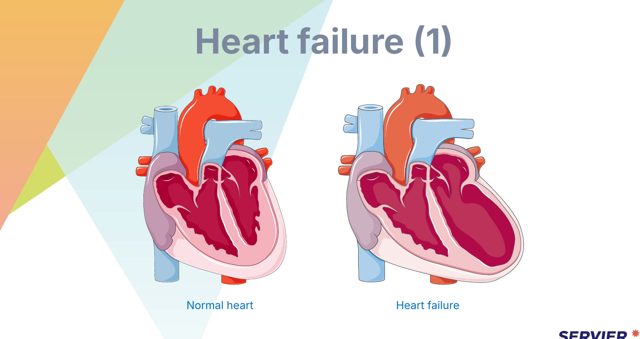
- So the signs are RESPIRATORY
- Exertional DYSPNOEA
- ORTHOPNOEA — measured in HOW MANY PILLOWS he needs
- PAROXYSMAL NOCTURNAL DYSPNOEA that wakes him
- CRACKLES, a cough with frothy or pink sputum, fatigue
- So the signs are SYSTEMIC
- RAISED jugular venous pressure
- PERIPHERAL OEDEMA — ankles, or over the SACRUM in a bed-bound patient
- HEPATOMEGALY, ascites, weight gain
- Nausea and anorexia from gut congestion
Two points that follow. The COMMONEST CAUSE of RIGHT-sided failure is LEFT-sided failure, so most patients eventually show BOTH. And right failure caused by LUNG disease rather than by the left heart is called COR PULMONALE — which is a distinction the exam asks for directly.
نقطتان تتبعان. أولاً: أشيع سبب لقصور القلب الأيمن هو قصور القلب الأيسر، فيُظهر معظم المرضى كليهما في النهاية. وثانياً: القصور الأيمن الناتج عن مرض رئوي لا عن القلب الأيسر يُسمّى القلب الرئوي — وهذا تمييز يسأل عنه الامتحان مباشرةً.
Right-sided failure backs up into the body, so the signs are systemic: raised jugular venous pressure, peripheral oedema in the ankles or over the sacrum in a bed-bound patient, hepatomegaly and right upper quadrant discomfort, ascites, weight gain and nausea from gut congestion.
القصور الأيمن يرتدّ إلى الجسم، فالعلامات جهازية: ارتفاع الضغط الوريدي الوداجي، ووذمة طرفية في الكاحلين أو فوق العجز لدى طريح الفراش، وضخامة كبد وانزعاج في الربع العلوي الأيمن، واستسقاء، وزيادة وزن وغثيان من احتقان الأمعاء.
The commonest cause of right-sided failure is left-sided failure, so most patients eventually show both. Right failure caused by lung disease rather than by the left heart is called cor pulmonale.
أشيع سبب للقصور الأيمن هو القصور الأيسر، فمعظم المرضى يُظهرون كليهما في النهاية. والقصور الأيمن الناجم عن مرض رئوي لا عن القلب الأيسر يُسمّى القلب الرئوي.
1.16 · Managing Heart Failure١٫١٦ · تدبير قصور القلب
Nursing management: position upright, oxygen if hypoxic, daily weight at the same time on the same scale after voiding, strict fluid balance, sodium and fluid restriction as prescribed, and pacing activity around fatigue as Chapter 06 of Part 01 sets out.
التدبير التمريضي: وضعية منتصبة، وأكسجين عند نقص الأكسجة، ووزن يومي في الوقت نفسه على الميزان نفسه بعد التبوّل، وميزان سوائل دقيق، وتقييد الصوديوم والسوائل كما هو موصوف، وتنظيم النشاط حول التعب كما يبيّن الفصل السادس من الجزء الأول.
The single most important thing to teach before discharge is daily weighing. A gain of more than about 1 kg in a day, or 2 to 2.5 kg in a week, is reported — because roughly 1 kg is 1 litre of retained fluid, and it appears days before the breathlessness that would otherwise bring him back to hospital.
أهمّ ما يُعلَّم قبل الخروج هو الوزن اليومي. فالزيادة فوق نحو كيلوغرام في اليوم، أو ٢ إلى ٢٫٥ كغم في الأسبوع، تُبلَّغ — لأن كيلوغراماً يعادل نحو لتر من السوائل المحتبسة، ويظهر قبل أيام من ضيق النفس الذي كان سيعيده للمستشفى.
Also teach: take the medication even when feeling well, do not stop a beta-blocker abruptly, report increasing breathlessness or needing more pillows, know which symptoms mean call for help today, keep immunisations up to date, and avoid non-steroidal anti-inflammatory drugs which cause fluid retention.
يُعلَّم أيضاً: تناول الدواء حتى عند الشعور بالتحسّن، وعدم إيقاف حاصر بيتا فجأةً، والإبلاغ عن تزايد ضيق النفس أو الحاجة لوسائد أكثر، ومعرفة الأعراض التي تعني طلب المساعدة اليوم، وتحديث التطعيمات، وتجنّب مضادّات الالتهاب غير الستيرويدية فهي تسبّب احتباس سوائل.
1.17 · Acute Pulmonary Oedema١٫١٧ · الوذمة الرئوية الحادّة
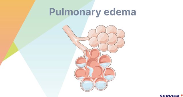
The picture is unmistakable: SEVERE breathlessness, EXTREME ANXIETY, coughing PINK FROTHY SPUTUM, WIDESPREAD CRACKLES, tachycardia, sweating, and a patient who CANNOT LIE FLAT.
الصورة لا لبس فيها: ضيق نفس شديد وقلق شديد وسعال ببلغم زهري رغوي وخراخر منتشرة وتسرّع قلب وتعرّق ومريض لا يستطيع الاستلقاء بوضع أفقي.
Position upright with the legs dependent — sitting up expands the lungs and letting the legs hang pools blood in them, reducing venous return and therefore preload. Give oxygen, obtain intravenous access, and expect an intravenous loop diuretic and nitrates; morphine has become less favoured than it once was.
يُوضَع المريض منتصباً والساقان متدلّيتان — فالجلوس يوسّع الرئتين وتدلّي الساقين يجمّع الدم فيهما، ما يقلّل العود الوريدي وبالتالي الحمل القبلي. ويُعطى الأكسجين، ويُؤمَّن مدخل وريدي، ويُتوقَّع مدرّ عروي وريدي ونترات؛ أما المورفين فصار أقلّ تفضيلاً ممّا كان.
Monitor continuously: oxygen saturation, respiratory rate, blood pressure, heart rhythm, and urine output, which tells you whether the diuretic is working and whether the kidneys are being perfused. Stay with the patient — the anxiety is part of the pathology and being left alone makes the breathlessness worse.
تجب المراقبة المستمرّة: تشبّع الأكسجين ومعدّل التنفّس وضغط الدم ونظم القلب وإدرار البول، فهو يبيّن هل يعمل المدرّ وهل تُروى الكليتان. ويلزم البقاء مع المريض — فالقلق جزء من المرض والترك وحيداً يزيد ضيق النفس.
1.18 · Atrial Fibrillation١٫١٨ · الرجفان الأذيني
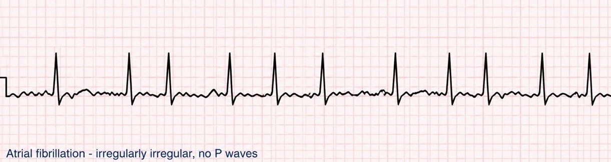
The most dangerous consequence is embolic stroke. The fibrillating atria do not empty properly, blood stagnates, a thrombus forms in the left atrial appendage and travels to the brain. That single fact is why anticoagulation, rather than rate control, is the intervention that changes outcomes.
أخطر عواقبه السكتة الدماغية الانصمامية. فالأذينان المرتجفان لا يُفرغان جيّداً، فيركد الدم، وتتشكّل خثرة في زائدة الأذين الأيسر وتنتقل للدماغ. وتلك الحقيقة وحدها هي سبب كون منع التخثّر، لا ضبط المعدّل، هو التدخّل الذي يغيّر المآل.
Management runs on three tracks: rate control with beta-blockers, calcium channel blockers or digoxin; rhythm control with drugs or cardioversion; and anticoagulation based on a stroke risk score. Before elective cardioversion the patient must be adequately anticoagulated, or the shock can dislodge an existing clot.
التدبير على ثلاثة مسارات: ضبط المعدّل بحاصرات بيتا أو قنوات الكالسيوم أو الديجوكسين؛ وضبط النظم بالأدوية أو تقويم النظم؛ ومنع التخثّر بناءً على درجة خطر السكتة الدماغية. وقبل تقويم النظم الاختياري يجب أن يكون المريض قد تلقّى مضادّات التخثّر بقدر كافٍ، وإلا فقد تُزيح الصدمة خثرةً موجودة.
1.19 · Grading Heart Failure and Oedema١٫١٩ · تدريج قصور القلب والوذمة
The New York Heart Association functional classification describes how much the symptoms limit the patient, and it is used to track change over time. Class I: no limitation of ordinary activity. Class II: slight limitation, comfortable at rest, ordinary activity causes symptoms. Class III: marked limitation, comfortable only at rest, less than ordinary activity causes symptoms. Class IV: symptoms at rest, and any activity worsens them.
التصنيف الوظيفي لجمعية القلب في نيويورك يصف مقدار تقييد الأعراض للمريض، ويُستخدم لتتبّع التغيّر مع الوقت. الصنف الأول: لا تقييد للنشاط الاعتيادي. والثاني: تقييد طفيف، مرتاح في الراحة، والنشاط الاعتيادي يُحدث أعراضاً. والثالث: تقييد ملحوظ، مرتاح في الراحة فقط، وأقلّ من النشاط الاعتيادي يُحدث أعراضاً. والرابع: أعراض في الراحة، وأيّ نشاط يزيدها.
Pitting oedema is graded by the depth of the indentation and how long it takes to rebound: 1+ a barely detectable pit of about 2 mm that disappears at once; 2+ about 4 mm, rebounding in a few seconds; 3+ about 6 mm, lasting longer; 4+ about 8 mm, a deep pit that remains for some time. Record where it was measured, since ankle oedema in an ambulant patient collects over the sacrum in someone confined to bed.
الوذمة الانطباعية تُدرَّج بعمق الانطباع وزمن ارتداده: +١ انطباع بالكاد يُلحَظ نحو ٢ ملم يزول فوراً؛ و+٢ نحو ٤ ملم يرتدّ خلال ثوانٍ؛ و+٣ نحو ٦ ملم يدوم أطول؛ و+٤ نحو ٨ ملم، انطباع عميق يبقى مدّة. ويُدوَّن موضع القياس، فوذمة الكاحلين لدى المريض المتنقّل تتجمّع فوق العجز لدى طريح الفراش.
1.20 · Bradycardia and Heart Block١٫٢٠ · بطء النبض وحصار القلب
Bradycardia matters when it is SYMPTOMATIC: hypotension, altered mental status, chest pain, acute heart failure, or signs of shock. A rate of 45 in an ASYMPTOMATIC athlete needs nothing — the number is read against the patient.
بطء القلب يهمّ حين يكون عرَضياً: هبوط ضغط أو تغيّر الحالة العقلية أو ألم صدر أو قصور قلب حادّ أو علامات صدمة. ومعدّل ٤٥ في رياضي بلا أعراض لا يحتاج شيئاً — فالرقم يُقرأ بحسب حال المريض.
The first-line drug is atropine 1 mg intravenously, repeated every 3 to 5 minutes to a maximum total of 3 mg. Note the dose: it was raised from 0.5 mg in the 2020 resuscitation guidance, so older materials give the earlier figure. If atropine fails, the next steps are transcutaneous pacing or an infusion of dopamine or adrenaline.
الدواء الأول أتروبين ١ ملغ وريدياً، يُكرَّر كلّ ٣ إلى ٥ دقائق حتى مجموع أقصاه ٣ ملغ. ويُلاحَظ في الجرعة أنها رُفعت من ٠٫٥ ملغ في إرشادات الإنعاش لعام ٢٠٢٠، فالمواد الأقدم تذكر الرقم السابق. وإن فشل الأتروبين فالخطوات التالية تنظيم ضربات عبر الجلد أو تسريب دوبامين أو أدرينالين.
Heart block in outline: first degree is a prolonged PR interval with every beat conducted; second degree Mobitz I shows a progressively lengthening PR until a beat drops; Mobitz II drops beats without warning and is more dangerous; third degree is complete dissociation of atria and ventricles and usually needs pacing.
حصار القلب باختصار: الدرجة الأولى إطالة فترة PR مع توصيل كلّ نبضة؛ والثانية موبيتز الأول إطالة تدريجية لـPR حتى تسقط نبضة؛ وموبيتز الثاني يُسقط نبضات دون إنذار وهو أخطر؛ والثالثة انفصال تامّ بين الأذينين والبطينين ويحتاج تنظيماً عادةً.
1.21 · Tachyarrhythmias and Cardioversion١٫٢١ · التسرّعات القلبية وتقويم النظم
Supraventricular tachycardia is a regular narrow-complex rhythm, typically 150 to 250 a minute, often starting and stopping abruptly. If the patient is stable, try vagal manoeuvres first — a Valsalva or carotid sinus massage where it is permitted. If those fail, adenosine 6 mg by rapid intravenous push followed immediately by a saline flush, and a further 12 mg if needed.
التسرّع فوق البطيني نظم منتظم ضيّق المركّب، ١٥٠ إلى ٢٥٠ في الدقيقة عادةً، ويبدأ وينتهي فجأةً غالباً. فإن كان المريض مستقرّاً، تُجرَّب المناورات المبهمية أولاً — مناورة فالسالفا أو تدليك الجيب السباتي حيث يُسمح. وإن فشلت، يُعطى أدينوزين ٦ ملغ بدفع وريدي سريع يتبعه فوراً غسل بمحلول ملحي، ثم ١٢ ملغ أخرى إن لزم.
Warn the patient before adenosine: its half-life is only seconds, and it commonly causes a brief but alarming flushing, chest tightness and a sense of impending doom, with a short pause on the monitor that is expected rather than a complication. Telling him in advance turns a frightening experience into a tolerable one.
يُحذَّر المريض قبل الأدينوزين: فعمر النصف ثوانٍ فقط، وهو يسبّب عادةً احمراراً في الوجه وضيقاً صدرياً وإحساساً بالهلاك الوشيك وجيزاً لكنه مفزع، مع توقّف قصير على الجهاز متوقّع لا مضاعفة. وإخباره مسبقاً يحوّل تجربة مخيفة إلى محتملة.
The distinction the exam tests: defibrillation is unsynchronised and is used for ventricular fibrillation and pulseless ventricular tachycardia. Synchronised cardioversion times the shock to the R wave and is used for an unstable tachycardia in a patient who still has a pulse — atrial fibrillation or flutter with haemodynamic compromise, SVT that has not responded, or ventricular tachycardia with a pulse. A conscious patient is sedated first.
التمييز الذي يختبره الامتحان: إزالة الرجفان غير متزامنة وتُستخدم للرجفان البطيني والتسرّع البطيني بلا نبض. وتقويم النظم المتزامن يُوقّت الصدمة على موجة R وتُستخدم لتسرّع غير مستقرّ لدى مريض ما زال له نبض — رجفان أو رفرفة أذينية مع اختلال في الدينامية الدموية، أو تسرّع فوق بطيني لم يستجب، أو تسرّع بطيني مع نبض. ويُهدَّأ المريض الواعي أولاً.
Atrial flutter produces a sawtooth baseline, often conducting two to one so the ventricular rate sits near 150. It carries the same embolic risk as atrial fibrillation and is anticoagulated on the same basis.
الرفرفة الأذينية تُنتج خطّ أساس منشاري الشكل، وتوصّل اثنين لواحد غالباً فيقع المعدّل البطيني قرب ١٥٠. وتحمل خطر الانصمام نفسه كالرجفان الأذيني ويُعطى فيها مضادّ التخثّر على الأساس نفسه.
1.22 · Cardiac Arrest and After١٫٢٢ · توقّف القلب وما بعده
High-quality compressions are the foundation: rate 100 to 120 per minute, depth 5 to 6 cm in an adult, full recoil between compressions, and minimal interruptions. For a shockable rhythm defibrillate immediately and resume compressions at once rather than pausing to check.
الضغطات عالية الجودة هي الأساس: معدّل ١٠٠ إلى ١٢٠ في الدقيقة، وعمق ٥ إلى ٦ سم لدى البالغ، وارتداد كامل بين الضغطات، وأقلّ انقطاع ممكن. وفي النظم القابل للصدمة تُجرى إزالة الرجفان فوراً وتُستأنف الضغطات مباشرةً بدل التوقّف للفحص.
Drugs: adrenaline 1 mg every 3 to 5 minutes, given as early as possible in a non-shockable rhythm and after the initial shocks have failed in a shockable one. Amiodarone is added for refractory ventricular fibrillation or pulseless ventricular tachycardia.
الأدوية: أدرينالين ١ ملغ كلّ ٣ إلى ٥ دقائق، يُعطى بأسرع ما يمكن في النظم غير القابل للصدمة، وبعد فشل الصدمات الأولى في القابل لها. والأميودارون يُضاف للرجفان البطيني المعند أو التسرّع البطيني بلا نبض.
Search for the reversible causes throughout, especially in a non-shockable rhythm: hypoxia, hypovolaemia, hydrogen ion (acidosis), hypo- or hyperkalaemia, hypothermia, and tension pneumothorax, tamponade, toxins, thrombosis coronary and thrombosis pulmonary. After return of circulation, comatose patients receive targeted temperature management at 32 to 37.5 °C for at least 36 hours, with fever actively prevented.
يُبحث عن الأسباب القابلة للعكس طوال الوقت، خاصّةً في النظم غير القابل للصدمة: نقص الأكسجة، ونقص الحجم، وأيون الهيدروجين (الحماض)، ونقص أو فرط البوتاسيوم، وانخفاض الحرارة، والاسترواح الضاغط، والاندحاس، والسموم، والخثار التاجي، والخثار الرئوي. وبعد عودة الدوران، يتلقّى المرضى الذين في غيبوبة تدبيراً حرارياً مستهدفاً عند ٣٢ إلى ٣٧٫٥ درجة مئوية لمدّة ٣٦ ساعة على الأقلّ، مع المنع الفعّال للحمّى.
1.23 · Pacemakers and Implantable Defibrillators١٫٢٣ · نواظم الخطى ومزيلات الرجفان المزروعة
- Strong ELECTROMAGNETIC FIELDS
- Arc welding, large running motors, industrial equipment
- MRI unless the device is MR-conditional and the protocol is followed
- Do not linger in security gates — show the card and request a hand search
- Microwave ovens
- Household appliances
- Mobile phones — held to the OPPOSITE ear and not carried in a breast pocket over the device
- Airport security walk-through, briefly
An IMPLANTABLE CARDIOVERTER DEFIBRILLATOR delivers a SHOCK for a dangerous rhythm. Teach that a SINGLE shock followed by FEELING WELL is REPORTED TO THE CLINIC, while MULTIPLE shocks, or a shock followed by feeling unwell, means EMERGENCY ATTENDANCE. And a person touching him during a shock may feel a tingle — it is not dangerous to them, which is worth telling the family so they do not let go of him.
مزيل الرجفان المزروع يُطلق صدمةً عند نظم خطير. ويُعلَّم المريض أن صدمة واحدة يتبعها شعور بالعافية تُبلَّغ للعيادة، بينما صدمات متعدّدة أو صدمة يتبعها توعّك تعني حضوراً إسعافياً. وقد يشعر من يلمس المريض أثناء الصدمة بوخز — ولا خطر عليه من ذلك، ويُستحسن إخبار الأسرة بهذا كي لا تتركه.
Teaching for home: check the pulse regularly and report a rate persistently below the set rate; carry the device identification card at all times; report dizziness, fainting, palpitations, prolonged hiccups — which can mean diaphragmatic stimulation — or swelling and redness at the site.
التعليم للمنزل: يُفحص النبض بانتظام ويُبلَّغ عن معدّل مستمرّ دون المعدّل المضبوط؛ وتُحمَل بطاقة تعريف الجهاز دائماً؛ ويُبلَّغ عن دوخة أو إغماء أو خفقان أو فواق مطوّل — وقد يعني تنبيه الحجاب — أو تورّم واحمرار في الموقع.
What to avoid and what is safe: keep away from strong electromagnetic fields — arc welding, large running motors, industrial equipment; hold a mobile phone to the opposite ear and keep it out of a breast pocket over the device; do not linger in airport security gates, show the card instead; and magnetic resonance imaging is avoided unless the device is MRI-conditional. Household appliances including microwave ovens are safe.
ما يُتجنّب وما هو آمن: الابتعاد عن الحقول الكهرومغناطيسية القوية — لحام القوس والمحرّكات الكبيرة العاملة والمعدّات الصناعية؛ ووضع الهاتف على الأذن المقابلة وإبقاؤه خارج جيب الصدر فوق الجهاز؛ وعدم الوقوف طويلاً عند بوّابات أمن المطار بل إظهار البطاقة؛ ويُتجنّب التصوير بالرنين المغناطيسي إلا إن كان الجهاز مشروطاً بالرنين. والأجهزة المنزلية بما فيها أفران الميكروويف آمنة.
1.24 · Pericarditis and Cardiac Tamponade١٫٢٤ · التهاب التامور واندحاس القلب
Pericarditis gives sharp, pleuritic chest pain that is worse lying flat and on inspiration and is relieved by sitting forward, with a pericardial friction rub and often widespread saddle-shaped ST elevation across the leads rather than in a single territory. That positional character helps to separate it from infarction at the bedside.
التهاب التامور يعطي ألماً صدرياً حادّاً جنبياً يزداد بالاستلقاء على الظهر وبالشهيق ويزول بالجلوس مع الميل للأمام، مع احتكاك تاموري وارتفاع ST منتشر سرجيّ الشكل عبر الاشتقاقات غالباً لا في منطقة واحدة. وهذه الصفة الوضعية تساعد على تمييزه من الاحتشاء عند السرير.
- SHARP, PLEURITIC chest pain
- WORSE lying FLAT and on INSPIRATION
- RELIEVED by SITTING FORWARD
- PERICARDIAL FRICTION RUB
- Often WIDESPREAD ST elevation — not confined to one territory, which distinguishes it from infarction
- Fluid in the pericardial sac COMPRESSING the heart so it CANNOT FILL
- BECK’S TRIAD: HYPOTENSION + MUFFLED heart sounds + DISTENDED neck veins
- Plus PULSUS PARADOXUS — an exaggerated fall in systolic on inspiration
- And tachycardia with a narrow pulse pressure
Tamponade is an OBSTRUCTIVE SHOCK and a MEDICAL EMERGENCY. The treatment is PERICARDIOCENTESIS to remove the fluid. Nursing: escalate IMMEDIATELY, give oxygen, obtain IV access, give fluid to maintain filling while awaiting drainage, monitor continuously, and prepare for the procedure — and note that fluid HELPS here, unlike in most other causes of a failing heart, because the problem is filling rather than pumping.
الاندحاس صدمة انسدادية وحالة إسعافية. والعلاج بزل التامور لتفريغ السائل. والتمريض: التصعيد فوراً، وإعطاء الأكسجين، وتأمين مدخل وريدي، وإعطاء السوائل للحفاظ على الامتلاء ريثما يتمّ التصريف، والمراقبة المستمرّة، والتحضير للإجراء — ويُلاحَظ أن السوائل تساعد هنا خلافاً لمعظم أسباب قصور القلب، لأن المشكلة في الامتلاء لا في الضخّ.
1.25 · Valvular Disease١٫٢٥ · أمراض الصمّامات
Aortic stenosis has a classic triad — angina, syncope and exertional dyspnoea — with a harsh systolic ejection murmur radiating to the carotids. Once symptoms appear the prognosis without intervention is poor, so a patient with aortic stenosis who begins to faint on exertion needs urgent referral rather than reassurance.
تضيّق الأبهر له ثلاثي كلاسيكي — ذبحة وإغماء وضيق نفس بالجهد — مع نفخة انقباضية قذفية خشنة تنتشر للسباتيين. وبمجرّد ظهور الأعراض يكون الإنذار سيئاً دون تدخّل، فالمريض بتضيّق أبهر الذي يبدأ بالإغماء عند الجهد يحتاج إحالة عاجلة لا طمأنة.
Mitral stenosis raises left atrial pressure, so it causes atrial fibrillation, pulmonary congestion and a risk of embolism. Mitral regurgitation gives a blowing systolic murmur at the apex radiating to the axilla, and an acute severe regurgitation after infarction is the papillary muscle rupture described earlier.
تضيّق التاجي يرفع ضغط الأذين الأيسر، فيسبّب رجفاناً أذينياً واحتقاناً رئوياً وخطر انصمام. وقصور التاجي يعطي نفخة انقباضية نافخة عند القمّة تنتشر للإبط، والقصور الشديد الحادّ بعد الاحتشاء هو تمزّق العضلة الحليمية المذكور سابقاً.
Patients with prosthetic valves need lifelong attention to two things: anticoagulation, with a higher INR target for a mechanical valve, and awareness of infective endocarditis — any unexplained fever in a patient with a prosthetic valve is investigated rather than treated as a simple infection.
مرضى الصمّامات الاصطناعية يحتاجون انتباهاً مدى الحياة لأمرين: العلاج بمضادّات التخثّر، بهدف أعلى للنسبة المعيارية الدولية (INR) في الصمّام الميكانيكي، والوعي بالتهاب الشغاف الخمجي — فأيّ حمّى غير مفسَّرة لدى مريض بصمّام اصطناعي تُستقصى ولا تُعالَج كعدوى بسيطة.
1.26 · Hypertension and Hypertensive Emergency١٫٢٦ · ارتفاع ضغط الدم وطارئ ارتفاع ضغط الدم
Most hypertension is asymptomatic, which is why it is called silent and why measurement technique matters — Part 01 Chapter 04 covers the errors that create a diagnosis that is not real. Teaching centres on adherence, salt reduction, weight, exercise, alcohol, smoking and home monitoring, and on the fact that treatment continues even when he feels well.
معظم حالات ارتفاع ضغط الدم بلا أعراض، ولهذا يُسمّى صامتاً ولهذا يهمّ أسلوب القياس — والفصل الرابع من الجزء الأول يغطّي الأخطاء التي تخلق تشخيصاً غير حقيقي. ويتمحور التعليم حول الالتزام وتقليل الملح والوزن والرياضة والكحول والتدخين والمراقبة المنزلية، وحول أن العلاج يستمرّ حتى عند الشعور بالعافية.
A hypertensive urgency is a very high pressure without evidence of acute organ damage. A hypertensive emergency is a very high pressure with acute damage — severe headache, visual disturbance, confusion or seizure, chest pain, breathlessness with pulmonary oedema, or a rising creatinine.
حالة ارتفاع ضغط الدم الملحّة هي ضغط مرتفع جداً دون دليل على تلف عضوي حادّ. وطارئ ارتفاع ضغط الدم هو ضغط مرتفع جداً مع تلف حادّ — صداع شديد أو اضطراب بصري أو تشوّش أو اختلاج أو ألم صدر أو ضيق نفس مع وذمة رئوية أو ارتفاع الكرياتينين.
In an emergency the pressure is lowered with intravenous agents in a monitored setting, and it is lowered gradually and in a controlled way rather than to normal quickly. Dropping it too fast causes cerebral, coronary and renal hypoperfusion — the organs have adapted to the higher pressure and a sudden fall causes a stroke rather than preventing one.
في الطارئ يُخفَّض الضغط بأدوية وريدية في بيئة مراقَبة، ويُخفَّض تدريجياً وبشكل مضبوط لا إلى الطبيعي بسرعة. فالخفض السريع يسبّب نقص تروية دماغية وتاجية وكلوية — إذ تكيّفت الأعضاء مع الضغط الأعلى، والهبوط المفاجئ يسبّب سكتة دماغية بدل أن يمنعها.
1.27 · Arterial and Venous Limb Disease١٫٢٧ · مرض الطرف الشرياني والوريدي
- INTERMITTENT CLAUDICATION — cramping pain on WALKING, relieved by REST
- REST PAIN worse when the leg is ELEVATED, relieved when it HANGS DOWN
- COLD, PALE, hairless limb; weak or absent pulses
- Ulcers on the TOES, HEEL and PRESSURE POINTS — PUNCHED-OUT, PAINFUL
- Slow capillary refill
- ACHING HEAVINESS worse on STANDING, relieved by ELEVATION
- WARM limb with OEDEMA
- BROWN HAEMOSIDERIN staining
- Ulcers around the MEDIAL MALLEOLUS — SHALLOW, IRREGULAR, WET
- Pulses PRESENT
Chronic venous insufficiency is the mirror image: aching heaviness that is worse on standing and relieved by elevation, a limb that is warm with oedema and brown haemosiderin staining, and ulcers that are shallow, irregular, weeping and on the medial malleolus.
القصور الوريدي المزمن صورة معكوسة: ثقل موجع يزداد بالوقوف ويزول بالرفع، وطرف دافئ بوذمة وتصبّغ بنّي بالهيموسيديرين، وقرح سطحية غير منتظمة نازّة على الكعب الإنسي.
The positioning follows directly and the exam tests it: in arterial disease keep the legs dependent so gravity helps the weak arterial flow reach the foot; in venous disease elevate the legs so gravity helps the blood return. Getting this backwards makes the patient worse.
الوضعية تتبع ذلك مباشرةً والامتحان يختبرها: ففي المرض الشرياني تُبقى الساقان متدلّيتين لتساعد الجاذبية الجريان الشرياني الضعيف على بلوغ القدم؛ وفي الوريدي تُرفع الساقان لتساعد الجاذبية عودة الدم. وعكس ذلك يزيد المريض سوءاً.
1.28 · Venous Thromboembolism and Aortic Emergencies١٫٢٨ · الانصمام الخثاري الوريدي وطوارئ الأبهر
Deep vein thrombosis presents with unilateral limb swelling, pain, warmth, redness and sometimes a palpable cord. Do not massage the limb. Escalate, expect anticoagulation, and know that the reason it matters is pulmonary embolism — sudden breathlessness, pleuritic chest pain, tachycardia, hypoxia and sometimes haemoptysis.
الخثار الوريدي العميق يتظاهر بتورّم أحادي الجانب وألم ودفء واحمرار وأحياناً حبل محسوس. لا يُدلَّك الطرف. يُصعَّد الأمر ويُتوقَّع العلاج بمضادّات التخثّر، ويُعلَم أن سبب أهمّيته الانصمام الرئوي — ضيق نفس مفاجئ وألم صدر جنبي وتسرّع نبض ونقص أكسجة ونفث دم أحياناً.
An abdominal aortic aneurysm may be found as a pulsatile abdominal mass, which is not palpated deeply or repeatedly. Rupture presents with sudden severe abdominal or back pain, hypotension and collapse, and it is an immediate surgical emergency.
أمّ الدم الأبهرية البطنية قد تُكتشف ككتلة بطنية نابضة، ولا تُجسّ بعمق ولا بتكرار. والتمزّق يتظاهر بألم بطني أو ظهري شديد مفاجئ وهبوط ضغط وانهيار، وهو طارئ جراحي فوري.
Aortic dissection gives sudden tearing or ripping pain, classically radiating to the back between the shoulder blades, often with a difference in blood pressure between the two arms and unequal pulses. Any patient with severe chest pain should have the blood pressure checked in both arms.
تسلّخ الأبهر يعطي ألماً مفاجئاً ممزّقاً، ينتشر كلاسيكياً للظهر بين لوحي الكتف، مع فرق في ضغط الدم بين الذراعين ونبضات غير متساوية غالباً. وأيّ مريض بألم صدر شديد ينبغي قياس ضغطه في الذراعين.
1.29 · Cardiac Surgery and Haemodynamic Monitoring١٫٢٩ · جراحة القلب ومراقبة الديناميكا الدموية
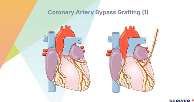
After coronary artery bypass grafting, sternal precautions protect the healing sternum: no lifting more than about 5 kg, no pushing or pulling with the arms — including pushing up out of a chair — and hugging a pillow against the chest when coughing, which allows an effective cough without splinting the pain away. If a leg vein was harvested, elevate that limb and watch the wound for infection.
بعد تطعيم مجازة الشريان التاجي، تحمي احتياطات القصّ عظم القصّ الملتئم: لا رفع فوق نحو ٥ كغم، ولا دفع أو شدّ بالذراعين — بما فيه الدفع للنهوض من كرسي — وضمّ وسادة للصدر عند السعال، ما يتيح سعالاً فعّالاً دون كبت الألم للسعال. وإن حُصد وريد من الساق فيُرفع ذلك الطرف ويُراقَب الجرح بحثاً عن عدوى.
Mediastinal and chest drains are measured hourly. Report drainage above roughly 100 to 150 mL an hour, and — the finding that is easily misread as good news — a sudden stop in drainage accompanied by hypotension, a rising central venous pressure and muffled heart sounds, which suggests the drain has clotted and blood is accumulating around the heart. That is tamponade. Atrial fibrillation is common in the days after surgery.
منازح المنصف والصدر تُقاس كلّ ساعة. يُبلَّغ عن تصريف يفوق نحو ١٠٠ إلى ١٥٠ مل في الساعة، وعن توقّف مفاجئ للتصريف — وهو الموجود الذي يسهل أن يُقرأ خبراً ساراً — مصحوب بهبوط ضغط وارتفاع الضغط الوريدي المركزي وأصوات قلب مكتومة، ما يوحي بتخثّر المنزح وتجمّع الدم حول القلب. وذلك اندحاس. والرجفان الأذيني شائع في الأيام التالية للجراحة.
Central venous pressure reflects right atrial filling and normally sits around 2 to 8 mmHg. A low value suggests hypovolaemia; a high value suggests fluid overload, right-sided failure or tamponade. Level and zero the transducer at the phlebostatic axis, and read the trend rather than a single figure.
الضغط الوريدي المركزي يعكس امتلاء الأذين الأيمن ويقع طبيعياً نحو ٢ إلى ٨ ملم زئبق. والقيمة المنخفضة توحي بنقص الحجم؛ والمرتفعة توحي بزيادة سوائل أو قصور أيمن أو اندحاس. ويُسوَّى المحوّل ويُصفَّر عند المحور الوريدي الساكن، ويُقرأ الاتّجاه لا رقم واحد.
Arterial line safety: perform an Allen test before radial insertion to confirm collateral flow; never inject anything into an arterial line; keep the alarms on and the connections visible and secure, because a disconnection causes rapid exsanguination; check the perfusion of the hand distal to it; and label the line clearly so it is not mistaken for a venous access.
سلامة الخطّ الشرياني: يُجرى اختبار ألن قبل التركيب الكعبري لتأكيد الجريان الجانبي؛ ولا يُحقَن أيّ شيء في خطّ شرياني أبداً؛ وتُبقى الإنذارات مفعّلة والوصلات مرئية ومحكمة، فالانفصال يسبّب نزفاً سريعاً مميتاً؛ وتُفحص تروية اليد بعده؛ ويُوسَم الخطّ بوضوح كي لا يُخلَط بمدخل وريدي.
For catheterisation by the radial rather than the femoral route, a compression band is applied at the wrist, the arm is kept straight, and the hand is checked for colour, warmth, sensation and movement. The radial approach carries less bleeding risk and allows earlier mobilisation.
للقسطرة عبر الطريق الكعبري بدل الفخذي، يُوضع رباط ضاغط عند الرسغ، وتُبقى الذراع مستقيمة، ويُفحص لون اليد ودفؤها وإحساسها وحركتها. والطريق الكعبري يحمل خطر نزف أقلّ ويتيح تحريكاً أبكر.
1.30 · Other Vascular Conditions١٫٣٠ · حالات وعائية أخرى
The ankle-brachial index compares the ankle systolic pressure with the brachial. A normal value is roughly 0.9 to 1.3; below 0.9 indicates peripheral arterial disease and below about 0.4 severe disease with critical ischaemia. A value above 1.3 suggests vessels too calcified to compress, which is common in diabetes and means the index cannot be relied on.
مشعر الكاحل العضدي يقارن ضغط الكاحل الانقباضي بالعضدي. والقيمة الطبيعية نحو ٠٫٩ إلى ١٫٣؛ ودون ٠٫٩ يدلّ على مرض شرايين طرفية ودون نحو ٠٫٤ على مرض شديد بنقص تروية حرج. والقيمة فوق ١٫٣ توحي بأوعية متكلّسة يتعذّر ضغطها، وهو شائع في السكّري ويعني أنه لا يمكن الاعتماد على المشعر.
Raynaud phenomenon is episodic vasospasm of the digits, classically producing a white then blue then red colour sequence, triggered by cold or emotional stress. Management is largely protective: keep the whole body and the hands warm, wear gloves including for the freezer at home, stop smoking, and avoid vasoconstricting drugs; calcium channel blockers are used where attacks are frequent.
ظاهرة رينو تشنّج وعائي نوبي في الأصابع، يُنتج كلاسيكياً تسلسل لون أبيض ثم أزرق ثم أحمر، ويُحدثه البرد أو الكرب الانفعالي. والتدبير وقائي أساساً: يُحافَظ على دفء الجسم كلّه واليدين، وتُلبَس قفّازات حتى عند التعامل مع الثلّاجة في المنزل، ويُقلَع عن التدخين، وتُتجنَّب الأدوية المقبّضة للأوعية؛ وتُستخدم حاصرات قنوات الكالسيوم حيث تتكرّر النوبات.
Varicose veins cause aching, heaviness and swelling that is worse on prolonged standing and relieved by elevation. Advise elevation when resting, graduated compression where it is not contraindicated, walking rather than standing still, avoiding crossing the legs, and skin care, since the skin over long-standing varicosities breaks down easily.
الدوالي تسبّب ألماً وثقلاً وتورّماً يزداد بالوقوف المطوّل ويزول بالرفع. ويُنصَح بالرفع عند الراحة، وبضغط متدرّج حيث لا مانع، وبالمشي بدل الوقوف ثابتاً، وبتجنّب تشبيك الساقين، وبالعناية بالجلد، فالجلد فوق الدوالي المزمنة يتلف بسهولة.
Carotid stenosis may be silent or produce a bruit, and it causes transient ischaemic attacks and stroke. A transient ischaemic attack — a neurological deficit that resolves completely — is a warning that must be acted on urgently rather than a reassuring event. Lymphoedema is swelling from impaired lymphatic drainage; it becomes non-pitting with thickened skin over time, and blood pressure measurement and venipuncture are avoided in the affected limb.
تضيّق السباتي قد يكون صامتاً أو يُنتج لغطاً، ويسبّب نوبات إقفارية عابرة وسكتة دماغية. والنوبة الإقفارية العابرة — عجز عصبي يزول تماماً — إنذار يجب التصرّف تجاهه عاجلاً لا حدث مطمئن. والوذمة اللمفية تورّم من ضعف التصريف اللمفي؛ وتصير غير انطباعية بجلد سميك مع الوقت، ويُتجنّب قياس الضغط والوخز الوريدي في الطرف المصاب.
1.31 · Shock١٫٣١ · الصدمة
SHOCK is INADEQUATE TISSUE PERFUSION, and it is classified by MECHANISM.
الصدمة تروية نسيجية غير كافية وتُصنَّف بحسب الآلية.
- TACHYCARDIA
- HYPOTENSION
- COOL, CLAMMY skin
- Prolonged capillary refill
- OLIGURIA and CONFUSION
- NEUROGENIC shock — HYPOTENSION with BRADYCARDIA
- and WARM, DRY, FLUSHED skin below the lesion
- EARLY SEPTIC shock — WARM peripheries and a BOUNDING pulse
- before it becomes cold and clamped down
- The exam relies on these two exceptions
The GENERAL priorities are the same whatever the cause: airway, breathing and circulation; oxygen; large-bore access; fluids WHERE VOLUME IS THE PROBLEM; and treating the cause. But the cause decides whether fluid helps or harms — which is why classifying it comes before treating it.
الأولويات العامّة واحدة أياً كان السبب: مجرى الهواء والتنفّس والدورة، والأكسجين، ومدخل وريدي واسع، والسوائل حيث تكون المشكلة في الحجم، وعلاج السبب. لكن السبب يحدّد هل تساعد السوائل أم تضرّ — ولهذا يسبق التصنيفُ العلاجَ.
Most shock looks the same at the bedside — tachycardia, hypotension, cool clammy skin, prolonged capillary refill, oliguria, confusion — but two types break that pattern and the exam relies on it. Neurogenic shock after a spinal cord injury above T6 gives bradycardia with hypotension and warm, dry, pink skin, because sympathetic tone is lost. Early septic shock can also give warm skin and a bounding pulse.
معظم الصدمات تبدو متشابهة عند السرير — تسرّع نبض وهبوط ضغط وجلد بارد رطب وتأخّر عود الشعيرات وقلّة بول وتشوّش — لكن نوعين يكسران هذا النمط والامتحان يعتمد عليه. فالصدمة العصبية بعد إصابة حبل شوكي فوق T6 تعطي بطء نبض مع هبوط ضغط وجلد دافئ جافّ وردي، لفقد المقوية الودّية. والصدمة الإنتانية المبكّرة قد تعطي جلداً دافئاً ونبضاً قافزاً.
The general priorities are the same whatever the cause: airway, breathing and circulation, oxygen, large-bore access, fluids where volume is the problem, and treating the cause. But the cause changes the treatment — fluids help hypovolaemia and can worsen cardiogenic shock, so identifying the mechanism is not academic.
الأولويات العامّة واحدة مهما كان السبب: مجرى الهواء والتنفّس والدورة، والأكسجين، ومدخل واسع، وسوائل حيث تكون المشكلة في الحجم، وعلاج السبب. لكن السبب يغيّر العلاج — فالسوائل تفيد نقص الحجم وقد تفاقم الصدمة القلبية، فتحديد الآلية ليس أمراً نظرياً.
1.32 · Cardiomyopathy, Rheumatic Disease and Prophylaxis١٫٣٢ · أمراض عضلة القلب، والمرض الروماتيزمي، والوقاية
Dilated cardiomyopathy — the ventricle enlarges and contracts poorly, giving systolic failure. Restrictive — the ventricle is stiff and fills poorly, giving diastolic failure. Hypertrophic — the muscle, especially the septum, thickens and can obstruct outflow; it is the classic cause of sudden cardiac death in a young athlete, so patients are advised to avoid strenuous competitive exercise and dehydration, and first-degree relatives are screened.
اعتلال عضلة القلب التوسّعي — يتّسع البطين وينقبض بضعف، فينشأ قصور انقباضي. والتقييدي — يتيبّس البطين ويمتلئ بضعف، فينشأ قصور انبساطي. والضخامي — تسمك العضلة وخاصّةً الحاجز وقد تسدّ المخرج؛ وهو السبب الكلاسيكي للموت القلبي المفاجئ لدى رياضي شابّ، فيُنصَح المرضى بتجنّب الرياضة التنافسية الشاقّة والجفاف، ويُفحَص أقارب الدرجة الأولى.
Rheumatic fever follows an untreated group A streptococcal throat infection, in which antibodies cross-react with cardiac tissue. It can leave permanent valve damage, most characteristically mitral stenosis. The point that matters for practice is that it is preventable: treating streptococcal pharyngitis with antibiotics prevents it, and patients who have had it receive long-term secondary prophylaxis to prevent recurrence.
الحمّى الروماتيزمية تتبع التهاب بلعوم بالعقدية من المجموعة أ غير معالَج، حيث تتفاعل الأضداد تصالبياً مع النسيج القلبي. وقد تترك تلفاً صمّامياً دائماً، وأكثره تميّزاً تضيّق التاجي. والنقطة المهمّة عملياً أنها قابلة للمنع: فعلاج التهاب البلعوم العقدي بالمضادات الحيوية يمنعها، ومن أصيبوا بها يتلقّون وقاية ثانوية طويلة الأمد لمنع النكس.
Infective endocarditis prophylaxis is now reserved for patients at the highest risk — a prosthetic valve, previous endocarditis, and certain congenital heart conditions — before dental procedures that involve manipulating the gums or perforating the oral mucosa. A single antibiotic dose is given before the procedure. For everyone else, good oral hygiene and regular dental care do more to prevent endocarditis than antibiotics do.
الوقاية من التهاب الشغاف الخمجي صارت مقصورة على الأعلى خطراً — صمّام اصطناعي، والتهاب شغاف سابق، وبعض حالات القلب الخلقية — قبل الإجراءات السنّية التي تشمل تحريك اللثة أو ثقب الغشاء المخاطي الفموي. وتُعطى جرعة واحدة من مضاد حيوي قبل الإجراء. أما لبقية الناس فنظافة الفم الجيّدة والعناية السنّية المنتظمة تمنع التهاب الشغاف أكثر ممّا تفعل المضادات الحيوية.
Amiodarone is effective but its adverse effects are wide-ranging and need monitoring: pulmonary toxicity and fibrosis, thyroid disturbance in either direction, hepatic impairment, corneal microdeposits, marked photosensitivity and a blue-grey skin discolouration, bradycardia and QT prolongation. Baseline and periodic thyroid function, liver function and chest imaging are the usual monitoring, and patients are told to use sun protection.
الأميودارون فعّال لكن آثاره الضارّة واسعة وتحتاج مراقبة: سمّية رئوية وتليّف، واضطراب درقي في أيّ اتّجاه، واختلال كبدي، ورواسب قرنية دقيقة، وحساسية ضوئية شديدة وتلوّن جلدي رمادي مزرقّ، وبطء نبض وإطالة QT. والمراقبة المعتادة وظيفة درقية وكبدية وتصوير صدر عند البدء ودورياً، ويُطلَب من المرضى استخدام الوقاية من الشمس.
1.33 · Antiplatelets and Anticoagulants١٫٣٣ · مضادّات الصفيحات ومضادّات التخثّر
Aspirin and the P2Y12 inhibitors such as clopidogrel are antiplatelets — they stop platelets aggregating and are the backbone of coronary disease treatment. In suspected infarction the aspirin is chewed for faster absorption. Watch for bleeding and gastrointestinal irritation.
الأسبرين ومثبّطات P2Y12 كالكلوبيدوغريل مضادّات صفيحات — توقف تكدّس الصفيحات وهي عماد علاج مرض الشرايين التاجية. وفي الاحتشاء المشتبه يُمضَغ الأسبرين لامتصاص أسرع. ويُراقَب النزف وتهيّج الجهاز الهضمي.
Heparin is monitored by the aPTT, targeted at roughly 1.5 to 2.5 times control, and its antidote is protamine sulfate. Watch the platelet count for heparin-induced thrombocytopenia. Low molecular weight heparin is given subcutaneously, needs less monitoring, and is not massaged after injection as Part 01 Chapter 07 sets out.
الهيبارين يُراقَب بزمن الثرومبوبلاستين الجزئي المفعّل، بهدف نحو ١٫٥ إلى ٢٫٥ ضعف الشاهد، وترياقه كبريتات البروتامين. ويُراقَب عدد الصفيحات تحسّباً لنقصها المحدَث بالهيبارين. والهيبارين منخفض الوزن الجزيئي يُعطى تحت الجلد ويحتاج مراقبة أقلّ ولا يُدلَّك بعد الحقن كما يبيّن الفصل السابع من الجزء الأول.
Warfarin is monitored by the INR, targeted at 2 to 3 for most indications and higher for a mechanical valve, and its antidote is vitamin K. Teach consistency rather than avoidance of vitamin K–rich foods, warn about the many drug interactions, and stress that the INR must be checked as scheduled.
الوارفارين يُراقَب بالنسبة المعيارية الدولية، بهدف ٢ إلى ٣ لمعظم الاستطبابات وأعلى للصمّام الميكانيكي، وترياقه فيتامين ك. ويُعلَّم الاتّساق لا تجنّب الأطعمة الغنية بفيتامين ك، ويُحذَّر من التداخلات الدوائية الكثيرة، ويُؤكَّد وجوب فحص النسبة في مواعيدها.
1.34 · Direct Oral Anticoagulants and Supportive Drugs١٫٣٤ · مضادّات التخثّر الفموية المباشرة والأدوية الداعمة
The direct oral anticoagulants — apixaban, rivaroxaban, edoxaban and dabigatran — differ from warfarin in ways that change nursing care. They need no routine INR monitoring, have far fewer food interactions, and act within hours. But their half-life is short, which means that missing doses leaves the patient unprotected quickly — adherence matters more, not less, than with warfarin. Doses are adjusted for renal function, age and weight, so renal monitoring is part of the care.
مضادّات التخثّر الفموية المباشرة — أبيكسابان وريفاروكسابان وإيدوكسابان ودابيغاتران — تختلف عن الوارفارين بما يغيّر الرعاية التمريضية. فهي لا تحتاج مراقبة روتينية للنسبة المعيارية الدولية (INR)، وتداخلاتها الغذائية أقلّ بكثير، وتعمل خلال ساعات. لكن عمر نصفها قصير، ما يعني أن إغفال الجرعات يترك المريض دون حماية سريعاً — فالالتزام يهمّ أكثر لا أقلّ ممّا مع الوارفارين. وتُعدَّل الجرعات لوظيفة الكلى والعمر والوزن، فالمراقبة الكلوية جزء من الرعاية.
Reversal differs too: idarucizumab reverses dabigatran, and andexanet alfa reverses apixaban and rivaroxaban. Vitamin K, which reverses warfarin, does not reverse these drugs — a distinction that matters in a bleeding patient, so establish which anticoagulant he is actually on rather than assuming.
عكس التأثير يختلف أيضاً: فالإيداروسيزوماب يعكس الدابيغاتران، والأندكسانيت ألفا يعكس الأبيكسابان والريفاروكسابان. وفيتامين ك الذي يعكس الوارفارين لا يعكس هذه الأدوية — وهو تمييز يهمّ لدى مريض ينزف، فيجب تحديد أيّ مضادّ تخثّر يتناوله فعلاً بدل الافتراض.
Potassium and diuretics: loop and thiazide diuretics cause potassium loss, so supplements or a potassium-sparing agent may be added. But potassium-sparing diuretics such as spironolactone, and ACE inhibitors and ARBs, all raise potassium — so a patient on those must not be given routine potassium supplements, and the level is checked before and during treatment. Combining several potassium-raising drugs is a recognised route to dangerous hyperkalaemia.
البوتاسيوم والمدرّات: المدرّات العروية والثيازيدية تسبّب فقد البوتاسيوم، فقد تُضاف مكمّلات أو عامل مُوفّر للبوتاسيوم. لكن المدرّات المُوفّرة للبوتاسيوم كالسبيرونولاكتون ومثبّطات الإنزيم المحوِّل للأنجيوتنسين وحاصرات مستقبل الأنجيوتنسين ترفع البوتاسيوم جميعاً — فلا تُعطى مكمّلات بوتاسيوم روتينية لمريض يتناول هذه الأدوية، ويُفحَص المستوى قبل العلاج وأثناءه. وجمع عدّة أدوية رافعة للبوتاسيوم طريق معروف لفرط بوتاسيوم خطير.
Sodium nitroprusside is a potent intravenous vasodilator used in hypertensive emergencies. It is light sensitive, so the bag and line are protected from light; it is given by infusion pump with continuous blood pressure monitoring; and prolonged or high-dose use risks cyanide and thiocyanate accumulation, so the duration is limited and the patient watched for confusion and metabolic acidosis.
نتروبروسيد الصوديوم موسّع وعائي وريدي قوي يُستخدم في طوارئ ارتفاع ضغط الدم. وهو حسّاس للضوء، فيُحمى الكيس والخطّ من الضوء؛ ويُعطى بمضخّة تسريب مع مراقبة ضغط مستمرّة؛ والاستخدام المطوّل أو بجرعة عالية ينطوي على خطر تراكم السيانيد والثيوسيانات، فتُحدَّد المدّة ويُراقَب المريض تحسّباً للتشوّش والحماض الاستقلابي.
1.35 · Preventing Venous Thromboembolism١٫٣٥ · الوقاية من الانصمام الخثاري الوريدي
Every patient is risk-assessed on admission and reassessed when the condition changes. The risk rises with surgery — orthopaedic especially — immobility, malignancy, previous clot, obesity, pregnancy, dehydration, oestrogen-containing medication and increasing age.
كلّ مريض يُقيَّم خطره عند القبول ويُعاد تقييمه عند تغيّر الحالة. والخطر يرتفع مع الجراحة — والعظمية خاصّةً — وقلّة الحركة والخباثة والخثرة السابقة والسمنة والحمل والجفاف والأدوية الحاوية للإستروجين وتقدّم العمر.
Mechanical prophylaxis: graduated compression stockings, correctly measured and fitted, and intermittent pneumatic compression devices. Stockings are contraindicated in peripheral arterial disease — compressing a limb whose arterial supply is already poor causes ischaemia — and are used with care where there is severe leg deformity, dermatitis or neuropathy. Check the skin underneath them daily.
الوقاية الميكانيكية: جوارب ضاغطة متدرّجة مقيسة ومركّبة بشكل صحيح، وأجهزة ضغط هوائي متقطّع. والجوارب ممنوعة في مرض الشرايين الطرفية — فضغط طرف إمداده الشرياني ضعيف أصلاً يسبّب نقص تروية — وتُستخدم بحذر حيث يوجد تشوّه ساق شديد أو التهاب جلد أو اعتلال عصبي. ويُفحَص الجلد تحتها يومياً.
Pharmacological prophylaxis is usually low molecular weight heparin, given subcutaneously and not massaged afterwards. Alongside both: early mobilisation, adequate hydration and leg exercises for anyone confined to bed. Homans sign is unreliable and is no longer used — it misses many clots and can dislodge one, so the diagnosis rests on the clinical picture and imaging.
الوقاية الدوائية هيبارين منخفض الوزن الجزيئي عادةً، يُعطى تحت الجلد ولا يُدلَّك بعده. وإلى جانب الاثنتين: تحريك مبكّر وإماهة كافية وتمارين للساقين لكلّ ملازم للفراش. وعلامة هومان غير موثوقة ولم تعد تُستخدم — فهي تُغفل خثرات كثيرة وقد تُزيح واحدة، فالتشخيص يقوم على الصورة السريرية والتصوير.
1.36 · Beta-Blockers, ACE Inhibitors and Calcium Channel Blockers١٫٣٦ · حاصرات بيتا ومثبّطات الإنزيم المحوِّل للأنجيوتنسين وحاصرات الكالسيوم
Beta-blockers slow the heart and lower blood pressure. Check the heart rate and blood pressure before every dose and hold if the rate is below about 60 or the systolic below about 90 to 100, then notify. Never stop them abruptly — rebound tachycardia, hypertension and ischaemia follow. They mask the warning signs of hypoglycaemia, which matters in a patient with diabetes, and are used cautiously in asthma.
حاصرات بيتا تبطئ القلب وتخفض الضغط. يُفحَص النبض والضغط قبل كلّ جرعة وتُحجَب الجرعة إن كان المعدّل دون نحو ٦٠ أو الانقباضي دون نحو ٩٠ إلى ١٠٠، ثم يُبلَّغ عن ذلك. ولا يُوقَف إعطاؤها فجأةً أبداً — فيتبع ذلك تسرّع نبض وارتفاع ضغط ونقص تروية ارتدادي. وهي تُخفي علامات إنذار نقص السكّر، وهذا يهمّ لدى مريض السكّري، وتُستخدم بحذر في الربو.
ACE inhibitors cause a persistent dry cough in a proportion of patients through bradykinin accumulation — this is an expected class effect, not an allergy, and the usual answer is to switch to an ARB. Watch also for angioedema, which is an emergency, hyperkalaemia, first-dose hypotension and rising creatinine. They are contraindicated in pregnancy.
مثبّطات الإنزيم المحوِّل للأنجيوتنسين تسبّب سعالاً جافّاً مستمرّاً لدى نسبة من المرضى بتراكم البراديكينين — وهو أثر متوقّع للصنف لا حساسية، والجواب المعتاد التحويل إلى حاصر مستقبل الأنجيوتنسين. ويُراقَب أيضاً الوذمة الوعائية وهي طارئ، وفرط البوتاسيوم، وهبوط ضغط الجرعة الأولى، وارتفاع الكرياتينين. وهي ممنوعة في الحمل.
Calcium channel blockers lower blood pressure and, in the case of the rate-limiting ones, slow the heart. Common effects are ankle oedema, flushing, headache and constipation. Grapefruit juice raises the levels of several of them, which is worth mentioning at discharge.
حاصرات قنوات الكالسيوم تخفض الضغط، والمُبطئة منها تبطئ القلب. والآثار الشائعة وذمة الكاحلين والتورّد والصداع والإمساك. وعصير الجريب فروت يرفع مستويات عدّة منها، ويستحقّ الذكر عند الخروج.
1.37 · Nitrates١٫٣٧ · النترات
NITRATES dilate VEINS and CORONARY ARTERIES, reducing PRELOAD and myocardial OXYGEN DEMAND. Check the BLOOD PRESSURE before EVERY dose and WITHHOLD if the systolic is below the specified threshold.
النترات توسّع الأوردة والشرايين التاجية فتقلّل الحمل القبلي وطلب العضلة القلبية للأكسجين. ويُفحَص ضغط الدم قبل كلّ جرعة وتُحجَب الجرعة إن كان الانقباضي دون الحدّ المحدَّد.
For sublingual use, teach the patient to sit or lie down first because of the hypotension and dizziness, place one tablet under the tongue, and if the pain is not relieved after 5 minutes, call the emergency number and then a further dose may be taken, up to three doses five minutes apart. Headache is common and expected. Tablets are kept in the original dark container and replaced as directed.
للاستخدام تحت اللسان، يُعلَّم المريض الجلوس أو الاستلقاء أولاً بسبب هبوط الضغط والدوخة، ووضع قرص واحد تحت اللسان، وإن لم يزل الألم بعد خمس دقائق فليتّصل برقم الطوارئ، ثم يجوز أخذ جرعة أخرى حتى ثلاث جرعات بفاصل خمس دقائق. والصداع شائع ومتوقّع. وتُحفظ الأقراص في عبوتها الداكنة الأصلية وتُستبدَل كما يُوجَّه.
Nitrates and phosphodiesterase-5 inhibitors must never be combined — the drugs used for erectile dysfunction such as sildenafil and tadalafil. Together they cause profound, refractory hypotension. Ask about them before giving a nitrate, and teach it explicitly at discharge, because a patient will not volunteer it.
لا يُجمَع بين النترات ومثبّطات الفوسفوديستراز-٥ أبداً — وهي أدوية ضعف الانتصاب كالسيلدينافيل والتادالافيل. فمعاً تسبّبان هبوط ضغط عميقاً معنداً. يُسأل عنها قبل إعطاء نترات، ويُعلَّم ذلك صراحةً عند الخروج، فالمريض لن يذكره تلقائياً.
1.38 · Digoxin, Diuretics and Statins١٫٣٨ · الديجوكسين والمدرّات والستاتينات
Digoxin slows the rate and strengthens contraction. Before every dose take the apical pulse for a full minute and hold if it is below 60 in an adult, then notify. The therapeutic range is narrow, so toxicity is common.
الديجوكسين يبطئ المعدّل ويقوّي الانقباض. وقبل كلّ جرعة يُؤخذ النبض القمّي دقيقةً كاملة وتُحجَب الجرعة إن كان دون ٦٠ لدى البالغ، ثم يُبلَّغ عن ذلك. والنطاق العلاجي ضيّق، فالسمّية شائعة.
Digoxin toxicity: anorexia, nausea and vomiting are usually first, then visual disturbance — classically yellow-green haloes or blurring — with confusion, bradycardia and arrhythmias. Hypokalaemia potentiates it, which is why a patient on both digoxin and a loop diuretic needs the potassium watched closely. Hold the drug, check the digoxin and potassium levels, and notify.
تسمّم الديجوكسين: فقد الشهيّة والغثيان والتقيّؤ أولاً عادةً، ثم اضطراب بصري — كلاسيكياً هالات صفراء خضراء أو زغللة — مع تشوّش وبطء نبض واضطراب نظم. ونقص البوتاسيوم يزيده، ولهذا يحتاج المريض على ديجوكسين ومدرّ عروي مراقبة لصيقة للبوتاسيوم. يُحجَب الدواء ويُفحَص مستوى الديجوكسين والبوتاسيوم ويُبلَّغ عن ذلك.
Loop diuretics such as furosemide cause hypokalaemia — so muscle weakness, cramps and arrhythmias — along with dehydration, hypotension and ototoxicity at high doses or rapid infusion. Give in the morning so the patient is not woken to pass urine, weigh daily, and monitor potassium. Statins: teach the patient to report unexplained muscle pain, tenderness or weakness immediately, since it can herald rhabdomyolysis.
المدرّات العروية كالفوروسيميد تسبّب نقص البوتاسيوم — فضعف عضلي وتشنّجات واضطراب نظم — مع جفاف وهبوط ضغط وسمّية أذنية بالجرعات العالية أو التسريب السريع. تُعطى صباحاً كي لا يُوقَظ المريض للتبوّل، ويُوزَن يومياً، ويُراقَب البوتاسيوم. والستاتينات: يُعلَّم المريض الإبلاغ فوراً عن ألم عضلي أو إيلام أو ضعف غير مفسَّر، فقد ينذر بانحلال الربيدات.
1.39 · Cardiovascular Care in the Saudi Setting١٫٣٩ · الرعاية القلبية الوعائية في السياق السعودي
Cardiovascular disease is a leading cause of death in the Kingdom, and it sits alongside a high prevalence of type 2 diabetes, obesity and physical inactivity. A Saudi study of adults with type 2 diabetes found established atherosclerotic cardiovascular disease in roughly one in seven, with coronary heart disease the commonest form. The practical consequence is that a Saudi adult with diabetes presenting with vague symptoms deserves a low threshold for a cardiac assessment.
أمراض القلب والأوعية من أبرز أسباب الوفاة في المملكة، وتقف إلى جانب انتشار مرتفع للسكّري من النمط الثاني والسمنة وقلّة النشاط البدني. ووجدت دراسة سعودية على بالغين بالسكّري من النمط الثاني مرضاً قلبياً وعائياً تصلّبياً مثبَتاً لدى نحو واحد من كلّ سبعة، وأشيع أشكاله مرض الشرايين التاجية. والنتيجة العملية أن البالغ السعودي المصاب بالسكّري الذي يحضر بأعراض غامضة يستحقّ عتبة منخفضة لتقييم قلبي.
Fasting in Ramadan is safe for most patients with stable cardiac disease, and the evidence does not show a rise in acute cardiac events during the month. Those advised not to fast include patients with unstable angina, a recent myocardial infarction, uncontrolled hypertension, decompensated heart failure, or recent cardiac intervention or surgery. The decision rests with the treating physician, and the Qur'an exempts the sick — a point worth stating gently, because many patients will fast regardless of advice.
الصيام في رمضان آمن لمعظم المرضى بمرض قلبي مستقرّ، والأدلّة لا تُظهر ارتفاعاً في الأحداث القلبية الحادّة خلال الشهر. ومن يُنصَحون بعدم الصيام مرضى الذبحة غير المستقرّة، والاحتشاء الحديث، وارتفاع ضغط الدم غير المضبوط، وقصور القلب غير المعوَّض، والتدخّل أو الجراحة القلبية الحديثة. والقرار للطبيب المعالج، والقرآن يُعفي المريض — وهي نقطة تستحقّ القول برفق، فكثير من المرضى سيصومون رغم النصيحة.
The nursing work in Ramadan is mostly about medication timing and fluid balance. Twice-daily regimens are often rescheduled to iftar and suhoor, and a switch to a once-daily preparation may be arranged in advance. Diuretic doses in particular may need adjusting, since a full dose during the fasting hours risks dehydration and hypotension; anticoagulated patients may see their INR fluctuate with the change in diet, so checks are kept up. Teach the warning symptoms that mean break the fast now — chest pain, severe breathlessness, dizziness or fainting.
العمل التمريضي في رمضان يدور أساساً حول توقيت الدواء وميزان السوائل. فالأنظمة مرّتين يومياً تُعاد جدولتها غالباً إلى الإفطار والسحور، وقد يُرتَّب مسبقاً التحويل إلى مستحضر مرّة واحدة يومياً. وجرعات المدرّات خاصّةً قد تحتاج تعديلاً، فالجرعة الكاملة خلال ساعات الصيام تنطوي على خطر الجفاف وهبوط الضغط؛ والمرضى الذين يتلقّون مضادّات التخثّر قد تتذبذب نسبتهم المعيارية مع تغيّر الحمية، فتُواصَل الفحوص. ويُعلَّم المريض الأعراض المنذرة التي تعني الإفطار الآن — ألم صدر أو ضيق نفس شديد أو دوخة أو إغماء.
Hajj and Umrah place REAL cardiovascular demands on pilgrims: sustained walking, dense crowds, heat and a disrupted routine.
الحجّ والعمرة يفرضان مطالب قلبية وعائية حقيقية على الحجّاج: المشي المتواصل، والازدحام الشديد، والحرارة، واضطراب الروتين.
Test yourselfالاختبار الذاتي
Loading… جارٍ التحميل…
Loading… جارٍ التحميل…
Loading… جارٍ التحميل…