
Basic Clinical Skills & Physiological Needs
In this chapterفي هذا الفصل
Key terms (12)المصطلحات الأساسية
Chapter outline (29 sections)مخطط الفصل
- Learning Objectivesأهداف التعلّم
- 7.1 · Positioning and What Each Position Achieves٧٫١ · الوضعيات وما تحقّقه كلّ منها
- 7.2 · Low-Flow Oxygen Devices٧٫٢ · أجهزة الأكسجين منخفضة التدفّق
- 7.3 · Reservoir and Controlled Devices٧٫٣ · أجهزة الخزّان والأجهزة المضبوطة
- 7.4 · Oxygen in the Patient at Risk of Retention٧٫٤ · الأكسجين لدى المريض المعرّض لاحتباس ثاني أكسيد الكربون
- 7.5 · Suctioning٧٫٥ · الشفط
- 7.6 · Tracheostomy Care٧٫٦ · العناية بفتحة القصبة
- 7.7 · Tonicity٧٫٧ · التوتّر
- 7.8 · Peripheral Intravenous Therapy٧٫٨ · العلاج الوريدي الطرفي
- 7.9 · Intravenous Complications٧٫٩ · مضاعفات العلاج الوريدي
- 7.10 · Blood Glucose and Insulin٧٫١٠ · سكّر الدم والإنسولين
- 7.11 · Eating, Dysphagia and Aspiration Risk٧٫١١ · الأكل وعسر البلع وخطر الاستنشاق
- 7.12 · Confirming Nasogastric Tube Placement٧٫١٢ · تأكيد موضع الأنبوب الأنفي المعدي
- 7.13 · Enteral Feeding٧٫١٣ · التغذية المعوية
- 7.14 · Parenteral Nutrition٧٫١٤ · التغذية الوريدية
- 7.15 · Urinary Elimination٧٫١٥ · الإخراج البولي
- 7.16 · Catheter Care and Infection Prevention٧٫١٦ · العناية بالقسطرة ومنع العدوى
- 7.17 · Bowel Elimination٧٫١٧ · الإخراج المعوي
- 7.18 · Enemas and Suppositories٧٫١٨ · الحقن الشرجية والتحاميل
- 7.19 · Stoma Care٧٫١٩ · العناية بالفغرة
- 7.20 · Staging a Pressure Injury٧٫٢٠ · تدريج قرحة الضغط
- 7.21 · Wound Healing and Assessment٧٫٢١ · التئام الجرح وتقييمه
- 7.22 · Dehiscence, Evisceration and Drains٧٫٢٢ · التفزّر والاندلاق والمنازح
- 7.23 · The Complications of Immobility٧٫٢٣ · مضاعفات قلّة الحركة
- 7.24 · Transfers and Walking Aids٧٫٢٤ · النقل ومعينات المشي
- 7.25 · Hygiene, Oral and Foot Care٧٫٢٥ · النظافة والعناية بالفم والقدم
- 7.26 · Sleep and Rest٧٫٢٦ · النوم والراحة
- 7.27 · Infusion Rates in Practice٧٫٢٧ · معدّلات التسريب عملياً
- 7.28 · Putting Physiological Needs in Order٧٫٢٨ · ترتيب الحاجات الفيزيولوجية
Start learningبدء التعلّم Learning objectivesأهداف التعلّم Test yourselfالاختبار الذاتي
Learning Objectivesأهداف التعلّم
7.1 · Positioning and What Each Position Achieves٧٫١ · الوضعيات وما تحقّقه كلّ منها
LOG ROLLING keeps the spine in ALIGNMENT as one unit, and needs enough people, with one person controlling the HEAD and giving the commands — used after spinal surgery and in any suspected spinal injury.
التدحرج الجذعي يُبقي العمود الفقري مصطفّاً كوحدة واحدة ويحتاج إلى عدد كافٍ من الأشخاص، يتحكّم أحدهم بالرأس ويعطي الأوامر؛ ويُستخدم بعد جراحة العمود الفقري وعند أيّ اشتباه بإصابة في العمود الفقري.
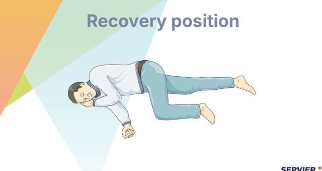
Orthopnoeic — sitting forward, arms resting on a table — lets the accessory muscles work and is what a severely breathless patient adopts on his own. Sims' — semi-prone on the left side with the upper knee flexed — is the position for an enema and for a rectal examination. The recovery position is for an unconscious patient who is breathing, and it protects the airway.
وضعية ضيق النفس — الجلوس مائلاً للأمام والذراعان على طاولة — تتيح للعضلات المساعدة العمل وهي ما يتّخذه المريض شديد ضيق النفس تلقائياً. ووضعية سيمز — شبه انبطاح على الجانب الأيسر مع ثني الركبة العليا — وضعية الحقنة الشرجية والفحص الشرجي. ووضعية الإفاقة لمريض فاقد الوعي يتنفّس، وهي تحمي مجرى الهواء.
Supine is flat on the back, prone face down, lateral on the side. Trendelenburg tips the head down and is used in specific circumstances rather than routinely for hypotension. Log rolling keeps the spine in alignment and needs enough people to turn the patient as one unit.
الاستلقاء الظهري مسطّحاً على الظهر، والانبطاح على الوجه، والجانبية على الجنب. ووضعية ترندلينبورغ تخفض الرأس وتُستخدم في ظروف محدّدة لا روتينياً لهبوط الضغط. والتدحرج الجذعي يُبقي العمود الفقري مصطفّاً ويحتاج عدداً كافياً من الأشخاص لتقليب المريض كوحدة واحدة.
7.2 · Low-Flow Oxygen Devices٧٫٢ · أجهزة الأكسجين منخفضة التدفّق
They are called LOW-FLOW devices because the flow they deliver is LESS THAN THE PATIENT’S INSPIRATORY DEMAND, so he ENTRAINS ROOM AIR alongside it. That is why the concentration they deliver is VARIABLE — it changes with his respiratory rate and depth. A breathless patient breathing hard gets a LOWER concentration from the same flow setting, which is the opposite of what people assume.
تُسمّى أجهزة منخفضة التدفّق لأن التدفّق الذي توصله أقلّ من طلب المريض الشهيقي فيسحب هواء الغرفة معه. ولهذا فالتركيز الذي توصله متغيّر، إذ يتغيّر بتغيّر معدّل تنفّس المريض وعمقه. والمريض الذي يعاني ضيق النفس ويتنفّس بقوّة يحصل على تركيز أقلّ من إعداد التدفّق نفسه، وهو عكس ما يظنّه الناس.
Simple face mask: roughly 5 to 10 litres per minute, delivering about 40 to 60%. The flow must never be set below 5 litres, because a lower flow does not clear the patient's exhaled carbon dioxide from the mask and he rebreathes it.
القناع الوجهي البسيط: نحو ٥ إلى ١٠ لترات في الدقيقة، ويعطي نحو ٤٠ إلى ٦٠٪. ولا يجوز ضبط التدفّق دون ٥ لترات أبداً، لأن التدفّق الأقلّ لا يطرد ثاني أكسيد الكربون المزفور من القناع فيعيد المريض استنشاقه.
These are called low-flow devices because the flow they deliver is less than the patient's inspiratory demand, so he entrains room air alongside it. That is why the concentration they deliver varies with his breathing pattern rather than being a fixed number.
تُسمّى أجهزة منخفضة التدفّق لأن التدفّق الذي تعطيه أقلّ من طلب المريض الشهيقي، فيسحب معه هواء الغرفة. ولهذا يتغيّر التركيز الذي تعطيه بحسب نمط تنفّسه ولا يكون رقماً ثابتاً.
7.3 · Reservoir and Controlled Devices٧٫٣ · أجهزة الخزّان والأجهزة المضبوطة
- 10–15 L/min → around 60–90% or more١٠–١٥ لتر/دقيقة ← نحو ٦٠–٩٠٪ أو أكثر
- The HIGHEST of any standard deviceالأعلى بين جميع الأجهزة القياسية
- ONE-WAY VALVES stop exhaled air entering the reservoirصمامات أحادية الاتجاه تمنع دخول الهواء المُزفَر إلى الخزّان
- INFLATE the reservoir bag BEFORE applying itيُنفخ كيس الخزّان قبل تطبيقه
- And it must never DEFLATE fully on inspiration — turn the flow upويجب ألّا يفرغ الكيس بالكامل عند الشهيق — يُرفع التدفّق
- 24, 28, 31, 35, 40 or 60%٢٤، ٢٨، ٣١، ٣٥، ٤٠ أو ٦٠٪
- Set by a COLOUR-CODED adaptor with a STATED FLOW for eachيُضبَط بمهايئ مُرمَّز بالألوان له تدفّق محدّد لكل نسبة
- Because it entrains a FIXED RATIO of airلأنه يسحب نسبة ثابتة من الهواء
- the concentration does NOT vary with his breathingلا يتغيّر التركيز مع نمط تنفّس المريض
- Which is why it is the device for a patient at risk of CO₂ retentionولهذا هو الجهاز المناسب لمريض معرّض لاحتباس ثاني أكسيد الكربون
HIGH-FLOW NASAL OXYGEN delivers HEATED, HUMIDIFIED gas at flows that EXCEED the patient’s inspiratory demand, giving a reliable concentration with better comfort. It sits between conventional oxygen and non-invasive ventilation — and it also provides a small amount of positive pressure and washes out dead space, which is why it does more than the concentration alone suggests.
الأكسجين الأنفي عالي التدفّق يوصل غازاً مسخَّناً مرطَّباً بتدفّقات تتجاوز طلب المريض الشهيقي فيعطي تركيزاً موثوقاً براحة أفضل. وهو يقع بين الأكسجين التقليدي والتهوية غير الباضعة، كما يوفّر قدراً صغيراً من الضغط الإيجابي ويغسل الحيّز الميت، ولهذا يفعل أكثر ممّا يوحي به التركيز وحده.
Venturi mask: delivers a precise, fixed percentage — commonly 24, 28, 31, 35, 40 or 60% — set by a colour-coded adaptor with a stated flow rate for each. Because it entrains a fixed ratio of room air, the concentration does not change with the patient's breathing, and that is exactly why it is the device of choice where the delivered percentage must be controlled.
قناع فنتوري: يعطي نسبة دقيقة ثابتة — عادةً ٢٤ أو ٢٨ أو ٣١ أو ٣٥ أو ٤٠ أو ٦٠٪ — تُضبط بمهايئ مُرمَّز بالألوان لكلّ منها معدّل تدفّق محدّد. ولأنه يسحب نسبة ثابتة من هواء الغرفة، لا يتغيّر التركيز بتنفّس المريض، وهذا بالضبط سبب كونه الجهاز المختار حيث يجب ضبط النسبة المعطاة.
High-flow nasal oxygen delivers heated, humidified gas at flows that exceed the patient's inspiratory demand, giving a reliable concentration with better comfort. It sits between conventional oxygen and non-invasive ventilation.
الأكسجين الأنفي عالي التدفّق يوصل غازاً مُسخَّناً مرطَّباً بتدفّقات تفوق طلب المريض الشهيقي، فيعطي تركيزاً موثوقاً براحة أفضل. وهو يقع بين الأكسجين التقليدي والتهوية غير الباضعة.
7.4 · Oxygen in the Patient at Risk of Retention٧٫٤ · الأكسجين لدى المريض المعرّض لاحتباس ثاني أكسيد الكربون
In a patient at risk of HYPERCAPNIC respiratory failure — chronic obstructive pulmonary disease is the usual example — uncontrolled HIGH-CONCENTRATION oxygen can WORSEN carbon dioxide retention. So a LOWER target saturation range is set and a CONTROLLED device (a Venturi mask) is used.
في مريض معرّض لقصور تنفّسي مترافق بفرط ثاني أكسيد الكربون — الانسداد الرئوي المزمن المثال المعتاد — قد يُفاقم الأكسجين عالي التركيز غير المضبوط احتباس ثاني أكسيد الكربون. لذلك يُحدَّد نطاق تشبّع مستهدف أدنى ويُستخدم جهاز مضبوط (قناع فنتوري).
The rule that must not be misapplied: a hypoxic patient is never left hypoxic for fear of retention. Oxygen is given and titrated to the target, and rising carbon dioxide is managed with monitoring and escalation, not by withholding oxygen from someone who is not oxygenating.
القاعدة التي يجب ألّا يُساء تطبيقها: المريض ناقص الأكسجة لا يُترك ناقص الأكسجة خوفاً من الاحتباس أبداً. فيُعطى الأكسجين ويُعاير للهدف، ويُدبَّر ارتفاع ثاني أكسيد الكربون بالمراقبة والتصعيد لا بحجب الأكسجين عمّن لا يتأكسج.
Watch for the signs of rising carbon dioxide: drowsiness, confusion, headache, flushed skin, a bounding pulse and a flapping tremor. A patient who becomes progressively harder to rouse after his oxygen was increased needs escalation and a blood gas, not simply more oxygen.
تُراقَب علامات ارتفاع ثاني أكسيد الكربون: النعاس والتشوّش والصداع وتورّد الجلد والنبض القافز والرعشة الخافقة. والمريض الذي يصير أصعب إيقاظاً تدريجياً بعد زيادة الأكسجين المعطى له يحتاج تصعيداً وغازات دم لا مزيداً من الأكسجين فحسب.
7.5 · Suctioning٧٫٥ · الشفط
Suction ONLY when there is a CLINICAL INDICATION — visible or audible secretions, a FALL IN SATURATION, coarse crackles, increased work of breathing, or a patient unable to clear his own secretions. Not routinely, and not to a schedule, because every pass causes harm as well as good.
يُشفَط فقط عند وجود استطباب سريري — إفرازات مرئية أو مسموعة أو هبوط في التشبّع أو خراخر خشنة أو زيادة جهد التنفّس أو عجز المريض عن تصفية إفرازاته بنفسه. ولا روتينياً ولا بجدول، لأن كلّ مرور يسبّب ضرراً إلى جانب الفائدة.
Technique: hyperoxygenate before and after; use sterile technique for tracheal and tracheostomy suction; insert without applying suction; apply suction only on withdrawal, rotating the catheter; and limit each pass to 10 to 15 seconds. Allow the patient to recover between passes and limit the number of passes.
الأسلوب: تُزاد الأكسجة قبل الشفط وبعده؛ وتُستخدم تقنية معقّمة لشفط الرغامى وفتحة القصبة؛ وتُدخَل القسطرة دون تطبيق الشفط؛ ويُطبَّق الشفط عند السحب فقط مع تدوير القسطرة؛ ويُقصَر كلّ مرور على ١٠ إلى ١٥ ثانية. ويُترك للمريض وقت للتعافي بين المرورات ويُحدّ من عدد المرورات.
Complications to watch for: hypoxia, cardiac arrhythmia including bradycardia from vagal stimulation, mucosal trauma and bleeding, raised intracranial pressure, bronchospasm, and infection. Stop and re-oxygenate if the saturation falls, the rhythm changes or the patient becomes distressed.
مضاعفات يجب مراقبتها: نقص الأكسجة، واضطراب النظم القلبي بما فيه بطء القلب من التنبيه المبهمي، ورضّ الغشاء المخاطي والنزف، وارتفاع الضغط داخل القحف، والتشنّج القصبي، والعدوى. يُتوقَّف عن الشفط وتُعاد الأكسجة إن انخفض التشبّع أو تغيّر النظم أو ظهرت على المريض علامات الكرب.
7.6 · Tracheostomy Care٧٫٦ · العناية بفتحة القصبة
Keep at the bedside at all times: a spare tube of the same size, a spare tube one size smaller, the obturator for the tube in place, a tracheal dilator where policy requires, suction equipment ready and working, and a bag-valve device with oxygen.
يُبقى عند السرير دائماً: أنبوب احتياطي بالمقاس نفسه، وأنبوب احتياطي بمقاس أصغر بدرجة واحدة، وسدّادة الإدخال (Obturator) الخاصة بالأنبوب الموجود، وموسّع رغامى حيث تشترط السياسة، ومعدّات شفط جاهزة تعمل، وجهاز كيس وصمّام مع أكسجين.
Routine care: humidification, because the upper airway that normally warms and moistens air has been bypassed; inner cannula care per policy; stoma site cleaning and inspection for skin breakdown; and tie changes performed by two people so the tube is held securely throughout.
الرعاية الروتينية: الترطيب، لأن المجرى العلوي الذي يدفّئ الهواء ويرطّبه قد جرى تجاوزه؛ والعناية بالقنية الداخلية وفق السياسة؛ وتنظيف موقع الفتحة وفحصه بحثاً عن تلف جلدي؛ وتغيير الأربطة بواسطة شخصين ليبقى الأنبوب ممسوكاً بإحكام طوال العملية.
Never use cut gauze around the stoma — loose fibres can be inhaled. If the tube becomes displaced or blocked, this is an airway emergency: call for help immediately, give oxygen over both the stoma and the mouth while help is coming, and follow the unit's emergency algorithm.
لا يُستخدم شاش مقصوص حول الفتحة أبداً — فالألياف السائبة قد تُستنشق. وإن انزاح الأنبوب أو انسدّ فهذه حالة طارئة في مجرى الهواء: تُستدعى المساعدة فوراً، ويُعطى الأكسجين على الفتحة والفم معاً ريثما تصل، وتُتّبع خوارزمية الطوارئ في الوحدة.
7.7 · Tonicity٧٫٧ · التوتّر
Hypotonic fluids such as 0.45% sodium chloride have a lower osmolality, so water moves into the cells. They are used for cellular dehydration and are avoided or used with great caution where cerebral oedema or raised intracranial pressure is a concern, because the brain swells too.
ناقصة التوتّر مثل كلوريد الصوديوم ٠٫٤٥٪ أسموليتها أقلّ، فينتقل الماء إلى الخلايا. وتُستخدم للجفاف الخلوي وتُتجنّب أو تُستخدم بحذر شديد حيث تثير الوذمة الدماغية أو ارتفاع الضغط داخل القحف القلق، فالدماغ ينتفخ أيضاً.
Hypertonic fluids such as 3% sodium chloride or 10% dextrose have a higher osmolality and pull water out of the cells into the vascular space. They expand circulating volume quickly and carry a real risk of fluid overload, so they are given with close monitoring.
مفرطة التوتّر مثل كلوريد الصوديوم ٣٪ أو الدكستروز ١٠٪ أسموليتها أعلى وتسحب الماء من الخلايا إلى الحيّز الوعائي. وتوسّع الحجم الدوراني بسرعة وتحمل خطراً حقيقياً بفرط حمل السوائل، فتُعطى بمراقبة لصيقة.
7.8 · Peripheral Intravenous Therapy٧٫٨ · العلاج الوريدي الطرفي
Choose the site with the plan in mind: a forearm vein is preferred; avoid areas of flexion, the side of a mastectomy, a limb with a fistula, an oedematous or paralysed limb, and the leg in an adult where it can be avoided. Use the smallest gauge that will do the job, and larger only where rapid volume or blood is needed.
يُختار الموقع مع وضع الخطة في الحسبان: يُفضَّل وريد الساعد؛ ويُتجنَّب كلٌّ من مناطق الثني وجهة استئصال الثدي وطرف به ناسور وطرف متوذّم أو مشلول والساق لدى البالغ حيث يمكن تجنّبها. ويُستخدم أصغر مقاس يؤدّي الغرض، والأكبر فقط حيث يلزم حجم سريع أو دم.
Change the administration set on the schedule your policy sets, not by habit. A set in continuous use does not need changing more often than every 96 hours, but is changed at least every 7 days.
يُبدَّل طقم الإعطاء بحسب الجدول الذي تحدّده السياسة المعتمدة لا بحكم العادة. والطقم المستعمل باستمرار لا يلزم تبديله أكثر من مرة كلّ ٩٦ ساعة، لكنه يُبدَّل مرة كلّ ٧ أيام على الأقل.
Inspect the site at every shift and before every infusion, and ask the patient whether it hurts — pain at the site is often the first sign of a problem and it comes before anything visible.
يُفحص الموقع كلّ مناوبة وقبل كلّ تسريب، ويُسأل المريض هل يؤلمه — فالألم في الموقع أول علامة على مشكلة غالباً ويسبق أيّ شيء مرئي.
7.9 · Intravenous Complications٧٫٩ · مضاعفات العلاج الوريدي
- A NON-VESICANT leaking into surrounding tissueتسرّب مادة غير منفِّطة إلى الأنسجة المحيطة
- Site is SWOLLEN, COOL and PALEالموضع متورّم وبارد وشاحب
- The infusion SLOWS; there may be discomfortيتباطأ التسريب؛ قد يكون هناك انزعاج
- STOP the infusion, REMOVE the cannulaيُوقَف التسريب وتُزال القُنيّة
- Elevate; apply warm or cold per the fluidيُرفع الطرف؛ وتُوضع كمادة دافئة أو باردة حسب نوع السائل
- Document and re-siteيُوثَّق ويُعاد التركيب في موضع آخر
- The SAME leakage but with a VESICANTالتسرّب نفسه لكن بمادة منفِّطة
- It can cause TISSUE NECROSISيمكن أن يسبّب نخر الأنسجة
- STOP immediatelyيُوقَف التسريب فوراً
- Do NOT remove the cannula until you have followed the policy — it may be needed to ASPIRATE or to give an ANTIDOTEلا تُزال القُنيّة قبل اتّباع السياسة — فقد يُحتاج إليها لسحب الدواء أو لإعطاء ترياق
- Escalate URGENTLY; mark, elevate, photograph, documentيُصعَّد الأمر بشكل عاجل؛ ويُعلَّم الموضع ويُرفع ويُصوَّر ويُوثَّق
PHLEBITIS is inflammation of the VEIN ITSELF. The site is RED, WARM and TENDER, often with a PALPABLE CORD along the vein’s course — which is the sign that distinguishes it. Stop the infusion, remove the cannula, apply warmth per policy, and re-site in the OTHER arm. And note the contrast that is examined: infiltration is COOL and PALE; phlebitis is WARM and RED.
التهاب الوريد التهاب في الوريد نفسه. فالموضع أحمر دافئ مؤلم مع حبل مجسوس غالباً على امتداد الوريد، وهو العلامة التي تميّزه. يُوقَف التسريب وتُنزع القنية ويُوضع الدفء وفق السياسة ويُعاد التركيب في الذراع الأخرى. ويُلاحَظ التباين الذي يُسأل عنه في الامتحان: أن التسرّب بارد شاحب والتهاب الوريد دافئ أحمر.
Extravasation is the same leakage but with a vesicant, and it can cause tissue necrosis. Stop immediately, do not remove the cannula until you have followed the policy — it may be needed to aspirate the drug — and escalate at once.
التسرّب خارج الوعاء هو التسرّب نفسه لكن بدواء منفِّط، وقد يسبّب نخر النسيج. يُوقَف التسريب فوراً، ولا تُنزع القنية قبل اتّباع السياسة — فقد تلزم لسحب الدواء — ويُصعَّد الأمر فوراً.
Phlebitis — inflammation of the vein itself. The site is red, warm and tender, often with a palpable cord along the vein's course. Stop the infusion, remove the cannula, apply warmth per policy, and document the grade. Circulatory overload presents with breathlessness, crackles, a raised jugular venous pressure and rising blood pressure: slow or stop the infusion, sit the patient up and escalate.
التهاب الوريد — التهاب في الوريد نفسه. فالموقع محمرّ ودافئ ومؤلم، مع حبل محسوس على مسار الوريد غالباً. يُوقَف التسريب وتُنزع القنية ويُوضع الدفء وفق السياسة وتُدوَّن الدرجة. وفرط الحمل الدوراني يتظاهر بضيق نفس وخراخر وارتفاع الضغط الوريدي الوداجي وارتفاع ضغط الدم: يُبطَّأ التسريب أو يُوقَف ويُجلَس المريض ويُصعَّد الأمر.
7.10 · Blood Glucose and Insulin٧٫١٠ · سكّر الدم والإنسولين
Insulin is drawn up with an insulin syringe only, and units are written in full — Chapter 05 covers why "U" is prohibited. For a single vial: inject air equal to the dose into the vial, then withdraw the dose.
يُسحب الإنسولين بمحقنة إنسولين فقط، وتُكتب الوحدات كاملةً — والفصل الخامس يبيّن لماذا حرف «U» ممنوع. وللقارورة الواحدة: يُحقن هواء بمقدار الجرعة في القارورة ثم تُسحب الجرعة.
When two insulins are mixed in one syringe the order matters, because contaminating the clear vial with the cloudy one alters its action. Inject air into the cloudy vial first, then air into the clear vial, then draw up the clear first and the cloudy second.
عند خلط نوعين من الإنسولين في محقنة واحدة يهمّ الترتيب، لأن تلويث القارورة الصافية بالعكرة يغيّر مفعولها. يُحقن الهواء في القارورة العكرة أولاً، ثم الهواء في الصافية، ثم تُسحب الصافية أولاً والعكرة ثانياً.
For a capillary blood glucose: wash and dry the hands with warm water rather than using alcohol which can alter the reading, sample from the side of the finger pad which is less painful and better perfused, avoid squeezing hard which dilutes the sample with tissue fluid, and rotate sites.
لسكّر الدم الشعري: تُغسل اليدان وتُجفَّفان بماء دافئ بدل استخدام الكحول الذي قد يغيّر القراءة، وتؤخذ العيّنة من جانب وسادة الإصبع فهو أقلّ إيلاماً وأفضل تروية، ويُتجنَّب العصر الشديد الذي يُخفّف العيّنة بسائل النسيج، وتُبدَّل المواقع.
7.11 · Eating, Dysphagia and Aspiration Risk٧٫١١ · الأكل وعسر البلع وخطر الاستنشاق
If dysphagia is suspected, keep the patient nil by mouth and refer for a swallowing assessment before giving food, fluid or oral medication. Silent aspiration — aspirating without coughing — is common after stroke and in neurological disease, and the absence of a cough is not reassurance.
إن اشتُبه بعسر البلع، يُبقى المريض صائماً ويُحال لتقييم البلع قبل إعطاء طعام أو سائل أو دواء فموي. والاستنشاق الصامت — الاستنشاق دون سعال — شائع بعد السكتة الدماغية وفي الأمراض العصبية، وغياب السعال ليس طمأنة.
Safe practice at mealtimes: sit the patient fully upright, keep the environment calm and unhurried, small mouthfuls, chin slightly tucked where advised, no talking with food in the mouth, check for pocketing, and keep him upright for at least 30 minutes afterwards. Provide oral care after eating.
ممارسة آمنة عند الوجبات: يُجلَس المريض منتصباً تماماً، وتُبقى البيئة هادئة غير مستعجلة، ولقيمات صغيرة، وذقن مطويّ قليلاً حيث يُنصح، ولا كلام والطعام في الفم، ويُتحقَّق من احتباس الطعام في الخدّ، ويُبقى منتصباً ثلاثين دقيقة على الأقلّ بعدها. وتُقدَّم عناية فموية بعد الأكل.
7.12 · Confirming Nasogastric Tube Placement٧٫١٢ · تأكيد موضع الأنبوب الأنفي المعدي
The accepted bedside method is testing the pH of aspirate: a pH in the acidic range, commonly 5.5 or below, is consistent with gastric placement. Acid-suppressing medication and feed can raise the pH, so an inconclusive result means re-checking or imaging, not proceeding.
الطريقة المقبولة عند السرير اختبار درجة حموضة الرشافة: فدرجة حموضة في المدى الحمضي، وعادةً ٥٫٥ أو أقلّ، تتّسق مع موضع معدي. وأدوية كبت الحموضة والتغذية ترفع درجة الحموضة (pH)، فالنتيجة غير الحاسمة تعني إعادة الفحص أو التصوير لا المضيّ.
Auscultating an air bolus is not a reliable method and is no longer recommended. A whoosh can be heard when the tube is in the lung, and patients have been fed into a lung on the strength of it. Also record and check the external length mark at every use, since a tube can migrate without any other sign.
الإصغاء لدفعة هواء ليس طريقة موثوقة ولم يعد موصى به. فقد يُسمع صوت اندفاع والأنبوب في الرئة، وقد غُذّي مرضى في رئاتهم اعتماداً عليه. ويُدوَّن كذلك ويُتحقَّق من علامة الطول الخارجي عند كلّ استخدام، فالأنبوب قد ينزاح دون أيّ علامة أخرى.
7.13 · Enteral Feeding٧٫١٣ · التغذية المعوية
Before every feed: confirm placement per policy, check the external length mark, and check gastric residual volume where the unit requires it. During and for at least 30 to 60 minutes after: keep the head of the bed at 30 to 45 degrees, which is the single most important measure against aspiration.
قبل كلّ تغذية: يُؤكَّد الموضع وفق السياسة، ويُتحقَّق من علامة الطول الخارجي، ويُفحص حجم البقايا المعدية حيث تشترطه الوحدة. وأثناءها ولثلاثين إلى ستّين دقيقة بعدها على الأقلّ: يُبقى رأس السرير بزاوية ٣٠ إلى ٤٥ درجة، وهو أهمّ تدبير منفرد ضدّ الاستنشاق.
Flush before and after the feed and between every medication, give each medication separately, use liquid formulations where available, and never add medication to the feed — Chapter 05 covers this in full. Some drugs bind to feed and require the feed to be paused around the dose.
يُغسل الأنبوب قبل التغذية وبعدها وبين كلّ دواء، ويُعطى كلّ دواء منفرداً، وتُستخدم الأشكال السائلة حيث تتوفّر، ولا يُضاف دواء إلى التغذية أبداً — والفصل الخامس يغطّي ذلك بالكامل. وبعض الأدوية ترتبط بالتغذية وتتطلّب إيقافها مؤقّتاً حول الجرعة.
Watch for and act on: aspiration, diarrhoea, constipation, nausea, abdominal distension, tube blockage and displacement, and skin excoriation at the nostril or stoma site. Provide mouth care even though the patient is not eating — a nil-by-mouth patient has a drier mouth, not a cleaner one.
يُراقَب ما يلي ويُتصرَّف حياله: الاستنشاق، والإسهال، والإمساك، والغثيان، وانتفاخ البطن، وانسداد الأنبوب وانزياحه، وتسلّخ الجلد عند المنخر أو موقع الفغرة. وتُقدَّم عناية فموية رغم أن المريض لا يأكل — فالصائم فمه أجفّ لا أنظف.
7.14 · Parenteral Nutrition٧٫١٤ · التغذية الوريدية
Parenteral nutrition is given when the gut cannot be used. It is usually delivered through a central line because of its high osmolality, and the lumen used is dedicated to it — no other medication, no blood sampling, no piggybacking.
تُعطى التغذية الوريدية حين يتعذّر استخدام الأمعاء. وتُقدَّم عبر قسطرة مركزية عادةً لأسموليتها العالية، ويكون التجويف المستخدم مخصّصاً لها — لا دواء آخر ولا سحب عيّنات ولا توصيل جانبي.
Never stop it abruptly. The patient's insulin response has adjusted to a high continuous glucose load, so a sudden stop causes rebound hypoglycaemia. It is tapered, and if a bag runs out before the next is available, a dextrose infusion is given per policy rather than nothing.
لا تُوقَف فجأةً أبداً. فاستجابة الإنسولين لدى المريض تكيّفت مع حمل غلوكوز مستمرّ عالٍ، فالإيقاف المفاجئ يسبّب هبوطاً ارتدادياً في سكّر الدم. وتُخفَّض تدريجياً، وإن نفد الكيس قبل توفّر التالي يُعطى تسريب دكستروز وفق السياسة بدلاً من عدم إعطاء أيّ شيء.
Monitor: blood glucose frequently, electrolytes, fluid balance and daily weight, temperature and the line site. Infection risk is high, so the line is handled with full aseptic technique at every access, as Chapter 05 sets out.
يُراقَب سكّر الدم بتواتر، والشوارد، وميزان السوائل والوزن اليومي، والحرارة وموقع القسطرة. وخطر العدوى مرتفع، فيُتعامل مع القسطرة بتقنية معقّمة كاملة عند كلّ استخدام كما يبيّن الفصل الخامس.
7.15 · Urinary Elimination٧٫١٥ · الإخراج البولي
Normal adult urine output is roughly 0.5 to 1 mL per kilogram per hour. A sustained output below 30 mL per hour in an adult is a warning sign of inadequate renal perfusion and is escalated rather than simply recorded. Oliguria is generally under 400 mL a day and anuria under 100 mL.
إدرار البول الطبيعي للبالغ نحو ٠٫٥ إلى ١ مل لكلّ كيلوغرام في الساعة. والإدرار المستمرّ دون ٣٠ مل في الساعة لدى بالغ علامة إنذار على تروية كلوية غير كافية ويُصعَّد الأمر ولا يُكتفى بتسجيله. وقلّة البول عموماً دون ٤٠٠ مل يومياً وانقطاع البول دون ١٠٠ مل.
Retention presents with a distended, palpable and often tender bladder, restlessness or agitation especially in an older patient, and sometimes small frequent volumes that are overflow rather than emptying. Confirm with a bladder scan where available before catheterising.
الاحتباس يتظاهر بمثانة متمدّدة محسوسة ومؤلمة غالباً، وتململ أو هياج خاصّةً لدى المسنّ، وأحياناً كمّيات صغيرة متكرّرة هي فيضان لا إفراغ. ويُؤكَّد ذلك بماسح مثانة حيث يتوفّر قبل القسطرة.
Types of incontinence worth distinguishing: stress on coughing or exertion, urge with a sudden need, overflow from a full bladder that does not empty, and functional where the bladder works but the patient cannot reach the toilet in time — which is often a mobility or environment problem the nurse can actually solve.
أنواع السلس الجديرة بالتمييز: الجهدي عند السعال أو الجهد، والإلحاحي بحاجة مفاجئة، والفيضاني من مثانة ممتلئة لا تُفرَغ، والوظيفي حيث تعمل المثانة لكن المريض لا يبلغ الحمّام في الوقت — وهو غالباً مشكلة حركة أو بيئة يستطيع الكادر التمريضي حلّها فعلاً.
7.16 · Catheter Care and Infection Prevention٧٫١٦ · العناية بالقسطرة ومنع العدوى
Insertion is a sterile procedure. Maintenance rests on four things: keep the system closed, keep the bag below bladder level so urine cannot flow back, keep the bag off the floor, and secure the catheter so it does not drag on the urethra.
التركيب إجراء معقّم. والصيانة تقوم على أربعة أمور: يُبقى النظام مغلقاً، ويُبقى الكيس تحت مستوى المثانة كي لا يرتجع البول، ويُبقى الكيس بعيداً عن الأرض، وتُثبَّت القسطرة كي لا تشدّ الإحليل.
The measure that outperforms all the others is reviewing the need daily and removing the catheter as soon as it is no longer indicated. Routine bladder irrigation and antiseptic meatal cleaning are not recommended and may increase risk; ordinary soap and water perineal hygiene is what is required.
التدبير الذي يتفوّق على غيره هو مراجعة الحاجة يومياً ونزع القسطرة فور انتفاء الاستطباب. والغسل المثاني الروتيني والتنظيف المطهّر للصماخ البولي غير موصى بهما وقد يزيدان الخطر؛ والمطلوب نظافة عجانية بماء وصابون عاديين.
Take a specimen from the sampling port after disinfecting it, never from the drainage bag, whose contents have been sitting and are not representative. Empty the bag using a separate clean container for each patient.
تؤخذ العيّنة من منفذ أخذ العيّنات بعد تطهيره، لا من كيس التصريف الذي ظلّ محتواه راكداً وغير ممثّل. ويُفرَغ الكيس بحاوية نظيفة منفصلة لكلّ مريض.
7.17 · Bowel Elimination٧٫١٧ · الإخراج المعوي
Assess the pattern rather than a fixed frequency — what is normal varies between people. Ask about consistency, effort, completeness, blood, and any change, since a change in bowel habit matters more than the habit itself.
يُقيَّم النمط لا التواتر الثابت — فما هو طبيعي يختلف بين الناس. ويُسأل عن القوام والجهد والاكتمال والدم وأيّ تغيّر، فتغيّر عادة الأمعاء أهمّ من العادة نفسها.
Constipation is managed by addressing the causes — fluid, fibre, mobility, privacy, opioids and other drugs, and ignoring the urge — before reaching for a laxative, and prophylactically in everyone on a regular opioid, as Chapter 06 sets out. Faecal impaction presents with a hard mass, abdominal discomfort, and often overflow diarrhoea that is mistaken for resolution.
الإمساك يُدبَّر بمعالجة الأسباب — السوائل والألياف والحركة والخصوصية والأفيونيات وأدوية أخرى وتجاهل الرغبة في التغوّط — قبل اللجوء لمليّن، ووقائياً لكلّ من يتناول أفيونياً بانتظام كما يبيّن الفصل السادس. والانحشار البرازي يتظاهر بكتلة صلبة وانزعاج بطني وغالباً بإسهال فيضاني يُظنّ خطأً أنه زوال للمشكلة.
For diarrhoea, assess hydration and electrolytes, protect the skin, and consider infection — a patient with new diarrhoea is isolated and stool sent before anyone reaches for an antidiarrhoeal, because suppressing an infective diarrhoea can be harmful. Chapter 05 covers the hand hygiene requirement.
في الإسهال، تُقيَّم الإماهة والشوارد، ويُحمى الجلد، ويُنظر في العدوى — فالمريض بإسهال جديد يُعزَل وتُرسَل عيّنة براز قبل أن يلجأ أحد لمضادّ إسهال، فكبت الإسهال الخمجي قد يضرّ. والفصل الخامس يغطّي اشتراط نظافة اليدين.
7.18 · Enemas and Suppositories٧٫١٨ · الحقن الشرجية والتحاميل
Position the patient in the left lateral Sims' position, because the sigmoid colon and descending colon lie on the left and the fluid then flows with gravity along the natural anatomy. Lubricate, insert gently along the direction of the rectum, and never force against resistance.
يُوضع المريض في وضعية سيمز الجانبية اليسرى، لأن القولون السيني والنازل يقعان يساراً فيسيل السائل مع الجاذبية على طول التشريح الطبيعي. ويُستعمل مزلّق، ويُدخَل برفق باتّجاه المستقيم، ولا يُدفع ضدّ مقاومة أبداً.
Use fluid at body temperature — too cold causes cramping and too hot injures the mucosa — and hang the solution low enough that it runs slowly, since height governs pressure. If the patient cramps, lower the container and pause rather than stopping altogether, then continue slowly.
يُستخدم سائل بحرارة الجسم — فالبارد جداً يسبّب مغصاً والساخن جداً يؤذي الغشاء المخاطي — ويُعلَّق المحلول منخفضاً بما يكفي ليجري ببطء، فالارتفاع يحكم الضغط. وإن أصاب المريض مغص، يُخفض الوعاء ويُتوقَّف مؤقّتاً بدل الإيقاف التامّ، ثم يُواصَل ببطء.
A cleansing enema is expelled; a retention enema is held for a stated period, so tell the patient which he is having and for how long. Ensure the call bell, a commode or the toilet are within reach before you begin, and give him privacy — dignity is part of the procedure, not an extra.
الحقنة المنظّفة تُطرَد؛ والمُحتبَسة تُحفظ مدّةً محدّدة، فيُخبَر المريض أيّ نوع سيتلقّاه ولكم من الوقت. ويُحرص على أن يكون جرس الاستدعاء وكرسي قضاء الحاجة (الكومود) أو المرحاض في المتناول قبل البدء، وتُمنح له خصوصية — فالكرامة جزء من الإجراء لا إضافة.
7.19 · Stoma Care٧٫١٩ · العناية بالفغرة
- PINK or RED, MOIST, slightly RAISEDوردي أو أحمر، رطب، مرتفع قليلاً
- NO nerve endings — so it is NOT painful to touchلا نهايات عصبية — لذا ليس مؤلماً عند اللمس
- Slight bleeding when cleaned is NORMAL — it is vascular tissueنزيف طفيف عند التنظيف أمر طبيعي — فهو نسيج وعائي
- It shrinks over the first weeks, so the appliance is re-measuredينكمش خلال الأسابيع الأولى، لذا يُعاد قياس الجهاز
- DUSKY, PURPLE, BLACK or DRY — ISCHAEMIAقاتم أو أرجواني أو أسود أو جافّ — نقص التروية
- This is the finding the exam asks about and the one that matters clinicallyهذه هي النتيجة التي يسأل عنها الاختبار وهي المهمّة سريرياً
- Retraction below skin levelتراجع الفغرة إلى ما تحت مستوى الجلد
- Prolapse, or a parastomal herniaتدلٍّ، أو فتق حول الفغرة
- And NO output with distension and pain — obstructionوانعدام الإخراج مع انتفاخ وألم — انسداد
A stoma that is dusky, purple, black or dry indicates ischaemia and must be reported immediately — this is the finding the exam asks about and the one that matters clinically. A pale stoma may reflect anaemia. Marked retraction, prolapse, separation from the skin or a stoma that stops working also need reporting.
الفغرة القاتمة أو الأرجوانية أو السوداء أو الجافّة تدلّ على نقص تروية ويجب الإبلاغ عنها فوراً — وهذه هي العلامة التي يسأل عنها الاختبار والتي تهمّ سريرياً. والفغرة الشاحبة قد تعكس فقر دم. والتراجع الشديد أو التدلّي أو الانفصال عن الجلد أو توقّف الفغرة عن العمل كلّها تحتاج إبلاغاً.
Protect the skin: cut the appliance to fit the stoma closely, since a gap lets output reach the skin. Ileostomy output is liquid, high in volume and corrosive to skin, and it carries a real risk of dehydration and electrolyte loss; colostomy output is more formed and less irritant. Measure and record the output.
يُحمى الجلد: يُقَصّ الجهاز ليطابق الفغرة بإحكام، فالفجوة تُوصل الصادر للجلد. وصادر الفغرة اللفائفية سائل وكبير الحجم وأكّال للجلد، ويحمل خطراً حقيقياً بالجفاف وفقد الشوارد؛ وصادر الفغرة القولونية أكثر تماسكاً وأقلّ تهيّجاً. ويُقاس الصادر ويُسجَّل.
7.20 · Staging a Pressure Injury٧٫٢٠ · تدريج قرحة الضغط
- The BASE is OBSCURED by SLOUGH or ESCHARالقاعدة مُغطّاة بنسيج نخري رخو أو جلبة
- So the DEPTH cannot be determinedلذا لا يمكن تحديد العمق
- The stage is assigned ONCE THE BASE IS VISIBLEتُحدَّد المرحلة بمجرد ظهور القاعدة
- It is at LEAST a stage 3 — you cannot obscure a stage 1 or 2هي على الأقل من المرحلة الثالثة — لا يمكن أن تُغطّى المرحلة الأولى أو الثانية
- PERSISTENT non-blanchable DEEP RED, MAROON or PURPLE discolourationتغيّر لون أحمر داكن أو عنّابي أو أرجواني مستمرّ لا يبيضّ
- Or a blood-filled blisterأو فقاعة مملوءة بالدم
- Damage has begun in the tissue BENEATH INTACT SKINالضرر بدأ في النسيج تحت الجلد السليم
- It can DETERIORATE RAPIDLY — and it looks minor from the surfaceيمكن أن يتدهور بسرعة — ويبدو بسيطاً من السطح
- Report it; do not wait to see what it becomesيُبلَّغ عنه؛ ولا يُنتظَر لمعرفة ما سيؤول إليه
Two rules that go with the staging: a pressure injury is NEVER ‘reverse-staged’ — a healing stage 4 does not become a stage 3; it is a healing stage 4. And MUCOSAL pressure injuries — from tubes, masks and devices — are NOT staged with this system at all, because the tissue is different.
قاعدتان مع التدريج: أن قرحة الضغط لا تُدرَّج عكسياً أبداً — فالمرحلة الرابعة الملتئمة لا تصبح ثالثة، بل تبقى رابعة ملتئمة؛ وإصابات الضغط المخاطية الناجمة عن الأنابيب والأقنعة والأجهزة لا تُدرَّج بهذا النظام إطلاقاً، لأن النسيج مختلف.
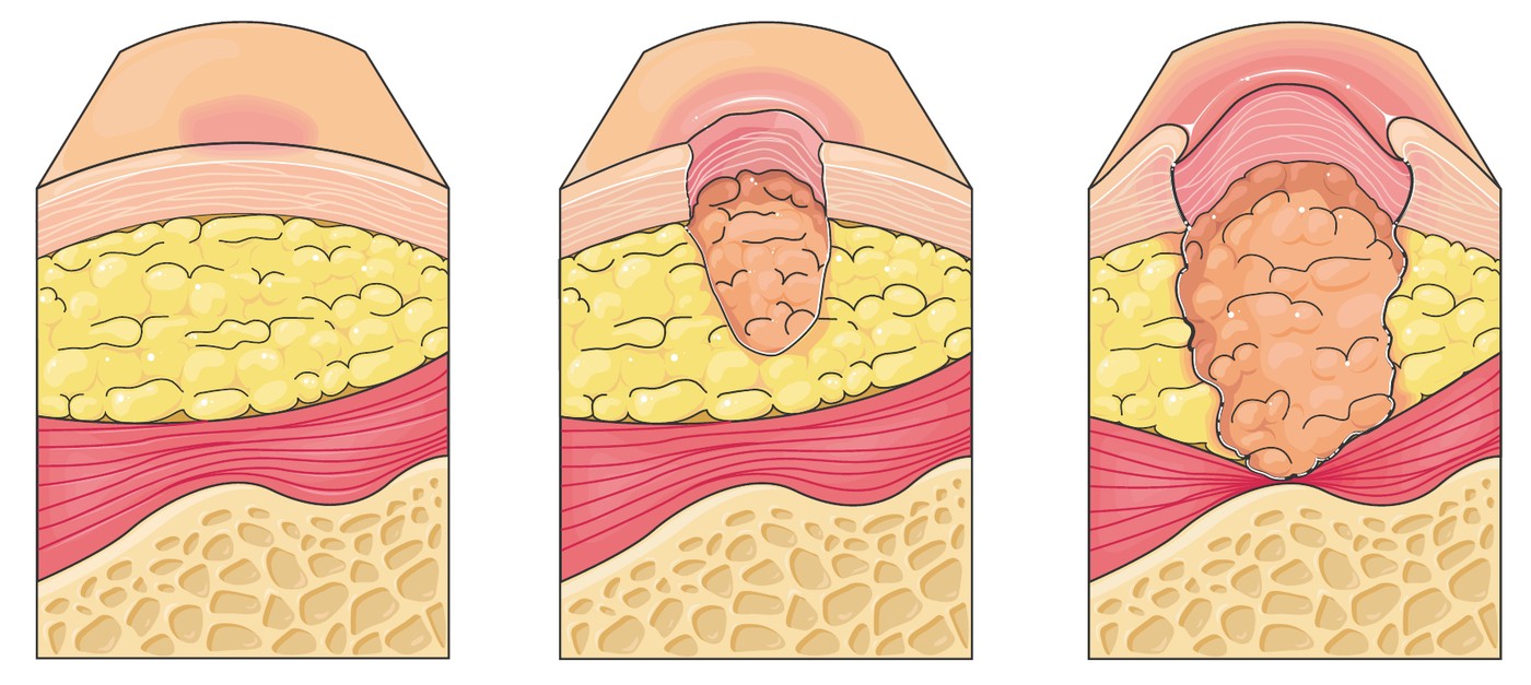
Stage 3: full-thickness loss in which subcutaneous fat is visible but bone, tendon and muscle are not exposed; slough may be present and there may be undermining or tunnelling. Stage 4: full-thickness loss with exposed bone, tendon or muscle.
المرحلة الثالثة: فقد كامل السماكة يظهر فيه الدهن تحت الجلد لكن العظم والوتر والعضل غير منكشفة؛ وقد يوجد نسيج نخري رخو وقد يوجد تقويض أو نفق. المرحلة الرابعة: فقد كامل السماكة مع انكشاف عظم أو وتر أو عضل.
Two categories that are not numbered. Unstageable: the base is obscured by slough or eschar so the depth cannot be determined — the stage is assigned once the base is visible. Deep tissue pressure injury: intact or non-intact skin with persistent, non-blanchable deep red, maroon or purple discolouration, or a blood-filled blister.
فئتان بلا رقم. غير القابلة للتدريج: القاعدة محجوبة بنسيج نخري رخو أو جلبة فيتعذّر تحديد العمق — وتُحدَّد المرحلة بمجرّد ظهور القاعدة. وإصابة الضغط في النسيج العميق: جلد سليم أو غير سليم مع تغيّر لون أحمر داكن أو عنّابي أو أرجواني مستمرّ لا يزول بالضغط، أو فقاعة مملوءة بالدم.
7.21 · Wound Healing and Assessment٧٫٢١ · التئام الجرح وتقييمه
Wounds close by primary intention — clean edges approximated, as with a surgical incision; secondary intention — left open to granulate from the base, as with a pressure injury; or tertiary intention, closed after a delay once contamination or oedema has settled.
تُغلق الجروح بالنيّة الأولى — حوافّ نظيفة متقاربة كالشقّ الجراحي؛ أو النيّة الثانية — تُترك مفتوحة لتتحبّب من القاعدة كقرحة الضغط؛ أو النيّة الثالثة، تُغلق بعد تأخير حين يزول التلوّث أو الوذمة.
Healing runs through haemostasis, an inflammatory phase in which redness, warmth and swelling are expected rather than abnormal, a proliferative phase of granulation and epithelialisation, and a long maturation phase in which the scar remodels and gains strength over months.
يمرّ الالتئام بالإرقاء، ثم مرحلة التهابية يكون فيها الاحمرار والدفء والتورّم متوقّعاً لا شاذّاً، ثم مرحلة تكاثرية من التحبّب والتظهّر، ثم مرحلة نضج طويلة يُعاد فيها تشكيل الندبة وتكتسب قوّة عبر أشهر.
Assess systematically: site, size, depth, undermining and tunnelling, the wound bed, exudate, odour, wound edges and the surrounding skin. The bed tells you a great deal — red granulation is healthy, yellow slough and black necrotic tissue are not. Photograph and measure so change can be judged rather than remembered.
يُقيَّم الجرح بشكل منهجي: الموقع والحجم والعمق والتقويض والنفق وقاع الجرح والنضح والرائحة وحوافّ الجرح والجلد المحيط. وقاع الجرح يدلّ على الكثير — فالتحبّب الأحمر سليم، أما النسيج النخري الأصفر والنسيج الأسود الميّت فلا. ويُصوَّر الجرح ويُقاس ليُحكَم على التغيّر لا ليُتذكَّر.
7.22 · Dehiscence, Evisceration and Drains٧٫٢٢ · التفزّر والاندلاق والمنازح
DEHISCENCE is SEPARATION of the wound layers. EVISCERATION is dehiscence WITH PROTRUSION OF ABDOMINAL CONTENTS — and it is a SURGICAL EMERGENCY. It often follows a sudden strain — coughing, vomiting, straining or sitting up — and is frequently preceded by a sudden GUSH of SEROSANGUINOUS fluid, which is the warning sign.
التفزّر انفصال طبقات الجرح. والاندلاق تفزّر مع بروز محتويات البطن وهو حالة إسعافية جراحية. وكثيراً ما يعقب جهداً مفاجئاً — سعالاً أو قيئاً أو إجهاداً أو جلوساً — ويسبقه غالباً خروج مفاجئ لسائل مصلي دموي، وهو علامة الإنذار.
DRAINS. A PENROSE drain is PASSIVE and drains onto a DRESSING. CLOSED SUCTION drains — a vacuum bottle or bulb device — are EMPTIED and then RE-COMPRESSED to RESTORE THE SUCTION, which is the step most often forgotten: an un-compressed bulb is a drain that has stopped working. Measure and record output each shift, keep the drain below the site, secure it against traction, and report a sudden increase, fresh blood, or a sudden STOP — because a drain that stops may be blocked rather than finished.
المنازح. منزح بنروز سلبي ويصرّف على ضمادة. ومنازح الشفط المغلقة — وهي جهاز زجاجة تفريغ أو بصلة — تُفرَغ ثم يُعاد ضغطها لاستعادة الشفط، وهي الخطوة الأكثر نسياناً: فالبصلة غير المضغوطة منزح توقّف عن العمل. ويُقاس الصادر ويُسجَّل كلّ مناوبة، ويُبقى المنزح دون مستوى الموضع، ويُثبَّت ضدّ الشدّ، ويُبلَّغ عن زيادة مفاجئة أو دم طازج أو توقّف مفاجئ — لأن المنزح الذي يتوقّف قد يكون مسدوداً لا منتهياً.
The actions, in order: stay with the patient and call for help; cover the protruding contents with sterile gauze moistened with sterile normal saline and keep it moist; position him low Fowler's with the knees bent to reduce tension on the abdomen; keep him nil by mouth; take observations and watch for shock; and notify the surgeon immediately.
الإجراءات بالترتيب: يُلازَم المريض وتُستدعى المساعدة؛ وتُغطّى المحتويات البارزة بشاش معقّم مبلّل بمحلول ملحي طبيعي معقّم ويُبقى رطباً؛ ويُوضع المريض في وضعية فاولر المنخفضة مع ثني الركبتين لتقليل الشدّ على البطن؛ ويُبقى صائماً؛ وتُؤخذ القياسات وتُراقَب علامات الصدمة؛ ويُبلَّغ الجرّاح فوراً.
Drains: a Penrose drain is passive and drains onto a dressing; closed suction drains such as a vacuum bottle or bulb device are emptied and then re-compressed to restore the suction, which is the step most often forgotten. Measure and record output separately for each drain, and report a sudden increase, a sudden stop, or a change to frank blood.
المنازح: منزح بنروز سلبي ويصرّف على ضماد؛ ومنازح الشفط المغلقة كزجاجة التفريغ أو جهاز البصلة تُفرَّغ ثم يُعاد ضغطها لاستعادة الشفط، وهي الخطوة الأكثر نسياناً. ويُقاس الصادر ويُسجَّل منفصلاً لكلّ منزح، ويُبلَّغ عن زيادة مفاجئة أو توقّف مفاجئ أو تحوّل إلى دم صريح.
7.23 · The Complications of Immobility٧٫٢٣ · مضاعفات قلّة الحركة
The single most effective intervention against NEARLY ALL of them is the SAME ONE: getting the patient MOVING, EARLY and REGULARLY. Not a different measure for each complication — one measure that addresses the respiratory, cardiovascular, musculoskeletal, gastrointestinal and psychological consequences at once.
أنجع تدخّل منفرد ضدّ جميعها تقريباً هو نفسه: تحريك المريض مبكّراً وبانتظام. فلا يلزم تدبير مختلف لكلّ مضاعفة، بل تدبير واحد يعالج العواقب التنفّسية والقلبية الوعائية والعضلية الهيكلية والهضمية والنفسية معاً.
Skin: pressure injury. Musculoskeletal: muscle atrophy, joint contractures, foot drop and bone demineralisation — prevented by range-of-motion exercises, correct positioning and support. Gastrointestinal: constipation and reduced appetite. Urinary: stasis, infection and calculi.
جلدياً: قرحة الضغط. وعضلياً هيكلياً: ضمور العضل وتقفّع المفاصل وسقوط القدم ونزع معادن العظم — ويُمنَع ذلك بتمارين مدى الحركة والوضعية الصحيحة والإسناد. وهضمياً: إمساك ونقص شهيّة. وبولياً: ركود وعدوى وحصيات.
Psychological: disorientation, low mood, anxiety and sleep disruption, which are easy to overlook and hard on the patient. The single most effective intervention against nearly all of these is getting the patient up and moving as early and as often as his condition allows.
نفسياً: اختلال التوجّه وانخفاض المزاج وقلق واضطراب نوم، وهي سهلة الإغفال وشاقّة على المريض. وأنجع تدخّل منفرد ضدّ جميعها تقريباً إنهاض المريض وتحريكه مبكّراً وبتكرار بقدر ما تسمح حاله.
7.24 · Transfers and Walking Aids٧٫٢٤ · النقل ومعينات المشي
A cane is held on the stronger side and advanced with the weaker leg, so that the cane and the weak leg share the load. A walking frame is advanced first, then the weaker leg, then the stronger — and the patient does not walk holding the frame at arm's length, which pulls him forward.
العصا تُمسَك في الجانب الأقوى وتتقدّم مع الساق الأضعف، لتتقاسم العصا والساق الضعيفة الحمل. والمشّاية تتقدّم أولاً، ثم الساق الأضعف، ثم الأقوى — ولا يمشي المريض ممسكاً بها على مدّ ذراعه، فذلك يجذبه للأمام.
If a patient begins to fall while you are with him, do not try to hold him upright. Widen your base, bring him close to your body and lower him slowly to the floor, protecting his head. Then assess him where he is before moving him, as Chapter 05 sets out.
إن بدأ مريض بالسقوط وهو برفقة مقدّم الرعاية، فلا يُحاوَل إبقاؤه واقفاً. تُوسَّع قاعدة الارتكاز، ويُقرَّب المريض من الجسم، ويُنزَل ببطء إلى الأرض مع حماية رأسه. ثم يُقيَّم في مكانه قبل تحريكه كما يبيّن الفصل الخامس.
7.25 · Hygiene, Oral and Foot Care٧٫٢٥ · النظافة والعناية بالفم والقدم
Hygiene is a clinical opportunity as much as a comfort measure: it is when the skin is inspected, mobility and pain are observed, and a conversation happens. Offer choice, maintain privacy and warmth, and work in a direction that avoids contaminating clean areas — perineal care is performed front to back.
النظافة فرصة سريرية بقدر ما هي تدبير راحة: فعندها يُفحَص الجلد وتُلاحَظ الحركة والألم ويحدث حديث. يُتاح للمريض الخيار، وتُحفظ الخصوصية والدفء، ويُعمل باتّجاه يتجنّب تلويث المناطق النظيفة — والعناية العجانية تُجرى من الأمام للخلف.
Oral care for an unconscious patient: position him side-lying with the head turned to the side, have suction ready and working, use minimal fluid, and never place fingers between the teeth. A dry mouth is a source of real discomfort, infection and thirst, and mouth care is one of the highest-value tasks on the ward.
العناية الفموية لمريض فاقد الوعي: يُوضع المريض على جانبه والرأس مُدار للجانب، ويُبقى الشفط جاهزاً يعمل، ويُستخدم أقلّ قدر من السوائل، ولا تُوضع الأصابع بين الأسنان أبداً. فجفاف الفم مصدر انزعاج حقيقي وعدوى وعطش، والعناية الفموية من أعلى المهامّ قيمةً في الجناح.
Diabetic foot care: inspect the feet daily including between the toes and the soles, wash and dry thoroughly especially between the toes, moisturise but not between the toes, cut nails straight across, never soak the feet, never use corn removers or sharp instruments, always wear something on the feet, and check the inside of shoes before putting them on.
العناية بقدم السكّري: تُفحص القدمان يومياً، ويشمل ذلك الأخمصين وما بين الأصابع، وتُغسلان وتُجفَّفان جيّداً خاصّةً بين الأصابع، ويُرطَّب الجلد لكن ليس بين الأصابع، وتُقَصّ الأظافر مستقيمةً، ولا تُنقع القدمان أبداً، ولا تُستخدم مزيلات مسامير القدم ولا أدوات حادّة، ويُلبَس شيء في القدمين دائماً، ويُتفقَّد داخل الحذاء قبل ارتدائه.
7.26 · Sleep and Rest٧٫٢٦ · النوم والراحة
Hospital is one of the worst environments for sleep: noise, light, interruptions for observations and medication, pain, anxiety, unfamiliar surroundings and daytime napping all disrupt it. Poor sleep worsens pain perception, mood, confusion and recovery, so it is a clinical problem rather than a comfort issue.
المستشفى من أسوأ البيئات للنوم: فالضجيج والضوء والمقاطعات للقياسات والأدوية والألم والقلق والمحيط غير المألوف والقيلولة النهارية كلّها تعطّله. وسوء النوم يفاقم إدراك الألم والمزاج والتشوّش والتعافي، فهو مشكلة سريرية لا مسألة راحة.
Nursing measures first: cluster care so the patient is not woken repeatedly, review whether overnight observations are still necessary, dim lights and reduce noise, control pain and offer toileting before settling, keep a consistent routine, avoid caffeine late, and encourage daytime activity and light.
التدابير التمريضية أولاً: تُجمع الرعاية كي لا يُوقَظ المريض مراراً، ويُراجَع مدى لزوم قياسات الليل، وتُخفَّض الإضاءة ويُقلَّل الضجيج، ويُضبَط الألم ويُعرَض قضاء الحاجة قبل الاستقرار للنوم، ويُحافَظ على روتين ثابت، ويُتجنَّب الكافيين متأخّراً، ويُشجَّع النشاط والتعرّض للضوء نهاراً.
A hypnotic is not the first response, particularly in an older adult, where sedatives raise the risk of falls, confusion and daytime drowsiness. If sleep remains poor after the environment and the pain have been addressed, that is the point to discuss it with the prescriber.
المنوّم ليس الاستجابة الأولى، خاصّةً لدى كبير السنّ، حيث ترفع المهدّئات خطر السقوط والتشوّش والنعاس النهاري. وإن بقي النوم سيّئاً بعد معالجة البيئة والألم، فتلك نقطة مناقشته مع الطبيب.
7.27 · Infusion Rates in Practice٧٫٢٧ · معدّلات التسريب عملياً
Drops per minute = volume in millilitres × the drop factor of the giving set ÷ the time in minutes. For 120 mL over 60 minutes with a set delivering 10 drops per mL: (120 × 10) ÷ 60 = 20 drops per minute.
القطرات في الدقيقة = الحجم بالمليلتر × عامل التقطير لطقم التسريب ÷ الزمن بالدقائق. فلـ١٢٠ مل خلال ٦٠ دقيقة بطقم تسريب يعطي ١٠ قطرات لكلّ مل: (١٢٠ × ١٠) ÷ ٦٠ = ٢٠ قطرة في الدقيقة.
Sense-check every answer before you act on it. Estimate first — 1,000 over 8 hours must be a bit more than 100 an hour — and if the number you calculate is far from your estimate, the arithmetic is wrong. Chapter 05 covers the dose formulas and the rule that an unusual volume or tablet count is a prompt to recheck.
تُراجَع معقولية كلّ جواب قبل التصرّف به. يُقدَّر الناتج أولاً — فـ١٠٠٠ خلال ٨ ساعات يجب أن يكون أكثر قليلاً من ١٠٠ في الساعة — وإن بعد الرقم المحسوب عن التقدير فالحساب خاطئ. والفصل الخامس يغطّي معادلات الجرعة وقاعدة أن الحجم أو عدد الأقراص غير المعتاد دعوة لإعادة التحقّق.
7.28 · Putting Physiological Needs in Order٧٫٢٨ · ترتيب الحاجات الفيزيولوجية
Applied to this chapter: a patient who cannot maintain his airway comes before a patient who is breathless, who comes before one who is hypotensive, who comes before an overdue feed or a routine wash. A blocked tracheostomy, an evisceration, a dusky stoma and a saturation that is falling are all immediate; a bed bath is not.
تطبيقاً على هذا الفصل: المريض الذي لا يستطيع الحفاظ على مجرى هوائه قبل المريض الذي يعاني ضيق النفس، وهو قبل من هبط ضغطه، وهو قبل تغذية متأخّرة أو استحمام روتيني. فانسداد فتحة القصبة والاندلاق والفغرة القاتمة والتشبّع الهابط كلّها فورية؛ أما الاستحمام بالسرير فلا.
The other half of prioritisation is what only you can do. Delegate what can safely be delegated within scope, as Chapter 01 sets out, and keep for yourself the assessment, the judgement and the escalation — those are the parts that cannot be handed over.
النصف الآخر من الترتيب هو ما لا يؤدّيه سوى المسؤول عن الرعاية. يُفوَّض ما يمكن تفويضه بأمان ضمن النطاق كما يبيّن الفصل الأول، ويُحتفظ بالتقييم والحكم والتصعيد لدى المسؤول عن الرعاية — فهي الأجزاء التي لا تُسلَّم.
Test yourselfالاختبار الذاتي
Loading… جارٍ التحميل…
Loading… جارٍ التحميل…
Loading… جارٍ التحميل…